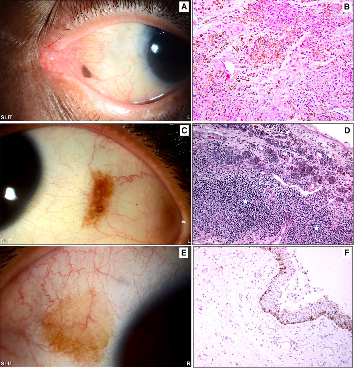
Fig. 4.
A The clinical photo of a small left eye nasal conjunctival pigmented lesion in an adult treated by excisional biopsy. B The histopathology of the same lesion showing melanocytic cells within the stroma with mild architectural atypia (loss of maturity) and no cytological atypia, finally diagnosed as a benign subepithelial nevus (original magnification × 200 hematoxylin and eosin). C The clinical appearance of a left eye temporal conjunctival pigmented lesion in a 15-year-old girl treated by excisional biopsy because of recent growth. D An example of the histopathological appearance of a conjunctival inflamed juvenile compound nevus (IJCN) with stromal lymphocytic infiltration (white stars) causing an increase in the nevus size clinically (original magnification × 200 periodic acid Schiff). E The clinical appearance of a lightly pigmented patch in a 30-year-old female that was excised without specific clinical diagnosis. F The histopathology of the conjunctival lesion revealed melanocytic hyperplasia without atypia, and the melanocytes at the base of the conjunctival epithelium expressed reactivity to the diagnostic immunohistochemical marker (original magnification × 100 Melan-A)

