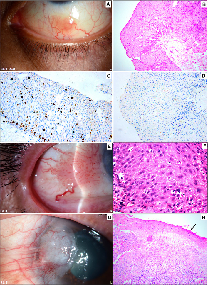
Fig. 5.
A Sessile gelatinous inferior conjunctival lesion in an adult female (33 years old) diagnosed clinically as a squamous papilloma. B The corresponding histopathological appearance of the papillomatous lesion with thick epithelium, absent dysplasia, and underlying fibrovascular core (original magnification × 200 hematoxylin and eosin). C The same lesion above showing a moderate proliferation index (original magnification × 200 Ki67). D The cells in the lesion did not show any reaction to human papilloma virus (HPV) antibodies, and a verrucous papilloma was excluded in this case (original magnification × 200 HPV). E Another sessile papillomatous temporal conjunctival lesion with a feeder blood vessel in a 72-year-old male treated by excisional biopsy with a clinical diagnosis of suspected OSSN. F The histopathological examination of a conjunctival lesion showing thick epithelium with moderate-to-severe dysplasia not involving the full thickness of the epithelium (original magnification × 400 hematoxylin and eosin). G The clinical appearance of a pterygium-like lesion extending into the cornea and showing evidence of leucoplakia. H Excised tissue showing histopathological evidence of invasive squamous cell carcinoma (SCC) with overlying metaplasia of the conjunctival epithelium and early keratinization (black arrow) corresponding to the area of leukoplakia (original magnification × 100 hematoxylin and eosin). Note the overlapping clinical appearance between a normal pterygium in Fig. 3A and the pterygium-like SCC in this latest case and the similarity of the sessile lesions in A and E that might lead to clinical misdiagnosis

