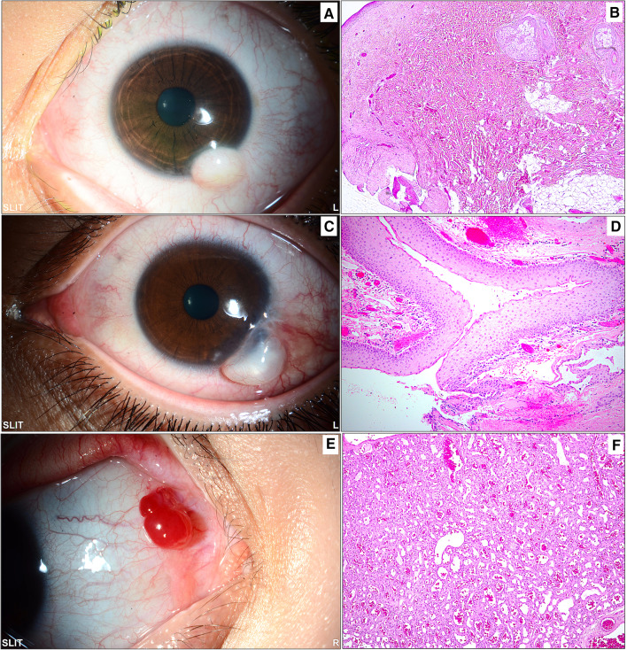
Fig. 7.
Miscellaneous lesions: A A whitish inferior dome-shaped limbal mass in a child diagnosed clinically as limbal dermoid. B The same lesion following excisional biopsy showing wavy thick collagenous fibers and fat with overlying skin-like epithelium and pilosebaceous units, confirming the diagnosis of a choristoma (original magnification × 50 hematoxylin and eosin). C The clinical appearance of a tumor-like cystic lesion in a 21-year-old male with long-standing severe vernal keratoconjunctivitis. D Histopathological picture of the collapsed cyst wall following excisional biopsy. The wall consists of nonkeratinizing stratified squamous epithelium representing the wall of an inclusion cyst (original magnification × 100 hematoxylin and eosin). E An example of a partially cystic red conjunctival lesion in a young female that was thought to be vascular in clinical nature. F The histopathology image showing lobular areas of capillary proliferation with the final diagnosis of a capillary hemangioma (original magnification × 100 hematoxylin and eosin)

