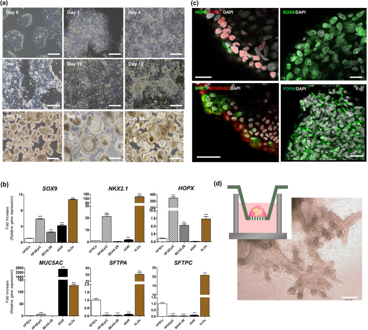
Fig. 1.
Generation of PSC-derived hLOs. a Bright-field images show the morphology of budding and differentiating 3D hLOs derived from PSCs in a time-dependent manner. Scale bar = 250 μm.) Lung-specific gene expression of the PSC-derived hLOs. The expression of SOX9, NKX2-1, HOPX, MUC5AC, SFTPA, and SFTPC in hLOs (brown bar). As controls, HPAEpiC cells (human primary alveolar epithelial cell line, hatched bar), BEAS-2B cells (human immortalized bronchial epithelial cell line, grey bar), and A549 cells (human lung cancer cell line, black bar) were used. The overall expression is relative to that in undifferentiated hPSCs on day 0. *p < 0.05, **p < 0.01, ***p < 0.001. All error bars represent SD. c Immunofluorescence data show that the hLOs express MUC1+ (green)/SCGB3A2+ (red): HOPX+ (green)/ NKX2-1+ (red): SOX9+ (green): PDPN+ (green) cells in the hLOs. The nucleus was visualized by DAPI staining (white). Scale bars = 20 μm. d Several new branches of hLOs in the Matrigel-coated Trans-well expanding over time were observed. Images of the hLOs on day 65 were captured by bright field microscopy. Scale bar = 200 μm

