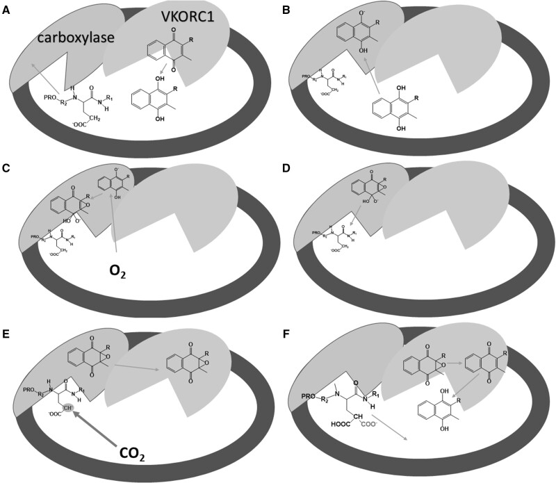
Figure 3.
Probable steps in the carboxylation process mediated by vitamin K in the endoplasmic reticulum. A protein that contains a PRO-sequence is targeted and subsequently bound to the carboxylase in the first step (A). This binding markedly increases the enzymatic function of the carboxylase. The quinone form of vitamin K is reduced to hydroquinone by VKORC1. Hydroquinone is deprotonated by carboxylase in the next step (B). Oxygen reacts with deprotonated vitamin K hydroquinone to produce alkoxide (C). This strong base deprotonates the γ-carbon of glutamyl residue to form a carbanion, which reacts with carbon dioxide (D–E). At the same time, vitamin K epoxide is formed (E). γ-glutamyl carboxylation is accomplished and the formed protein is released from the enzyme and further transported to the Golgi apparatus (not shown), while vitamin K epoxide is converted first to vitamin K quinone and then to vitamin K hydroquinone (F) by VKORC1. Data for this figure were taken from Rishavy et al (2004),75 Down et al (1995),76 Ayombil et al (2020)77 and Berkner (2000)78Abbreviations: carboxylase, vitamin K–dependent γ-glutamyl carboxylase; VKORC1, vitamin K epoxide reductase

