Figure 5.
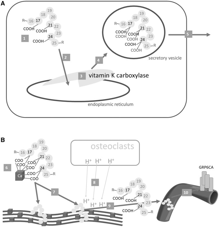
Osteocalcin. A: Synthesis and release from the osteoblast. B: Effect on bones and systemic release. Osteocalcin (1) is synthesized as preproprotein, then travels to the endoplasmic reticulum (2), where 3 glutamic acid residues at positions 17, 21 and 24 are γ-carboxylated by γ-glutamyl carboxylase (vitamin K carboxylase) (3). The carboxylated osteocalcin is further processed (eg, see Figure 4), transported in the vesicles (4), and released into the bone matrix (5), where it binds calcium ions in hydroxyapatite (6). This is a crucial step in bone formation – the correct alignment of collagen fibers with hydroxyapatite (7). When osteoresorption takes place, osteoclasts decrease the pH level in the bones (8); this can lead to the decarboxylation of osteocalcin to form un(der)carboxylated osteocalcin, which is released into the systemic circulation (9), where it could exert its hormonal effect, in particular by binding to the GRP6CA receptor (10)

