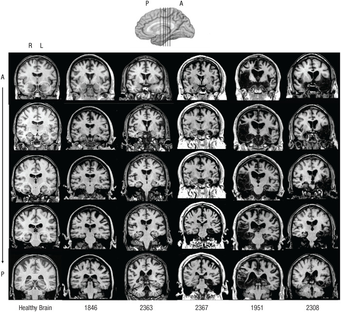
Fig. 1.
Magnetic resonance images of five amnesic patients and a normal healthy comparison brain. For each amnesic patient, a series of coronal MRI slices shows damage to the medial temporal lobes following anoxia (Patients 1846 and 2363), stroke (Patient 2367), or herpes simplex encephalitis (Patients 1951 and 2308). (Patients 2563 and 3139 could not undergo MRI because of the presence of a pacemaker.) R = right; L = left; A = anterior; P = posterior.

