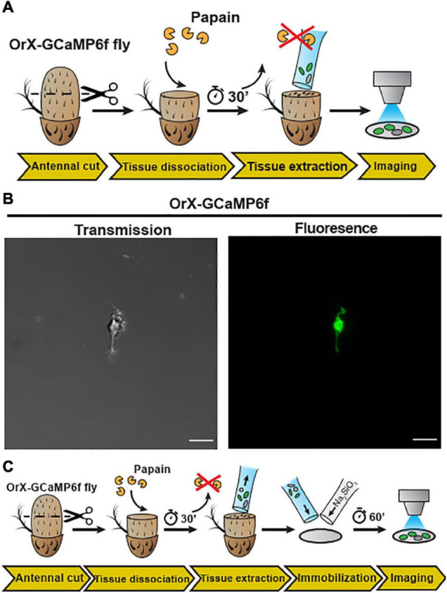
FIGURE 2.

Embedding of dissociated D. melanogaster antennal tissue in a sodium metasilicate gel. (A) Schematic procedure for dissociation and embedding of vinegar fly antennal tissue (see section “Materials and Methods”). After fixing the antenna with a silicon-based curing medium, the funiculus was cut and incubated with a papain solution for 30 min. Then, the dissociated tissue was extracted with a silanized glass capillary and then single neuron was extracted from this fly (+; Orco-Gal4/CyO; UAS-Syn21-GFP-p10/TM6B), using this preparation. (B) Confocal image of single cell as shown in transmission (gray) and fluorescence (green) signals, Scale bar = 8 μm. In the next illustration (C), after tissue extraction, the sample was mixed with a modified Drosophila Schneider’s medium containing 0.972% of a Na2SiO3 stock solution (≥27% SiO2 basis) on a methanol treated cover slip. Ca2+ imaging was performed after a 60 min incubation time, to allow the Na2SiO3 gelation.
