Figure 2.
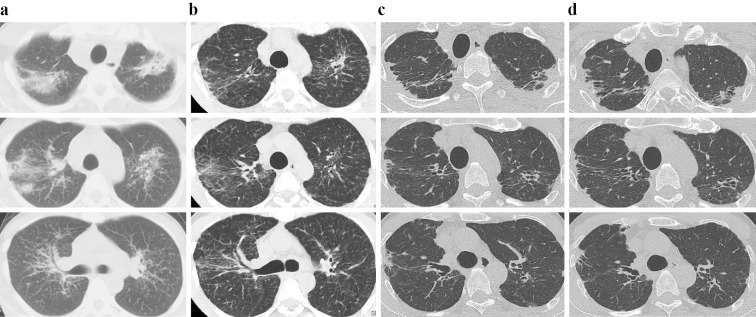
Chest CT images. (a) In 2004, typical early sarcoidosis lesions in the lung field (i.e. multiple micronodular and infiltrative opacities around the bronchovascular bundle in both upper lobes) and bilateral hilar and mediastinal lymphadenopathy were observed. (b) In 2006, the micronodular and infiltrative opacities had decreased bilaterally in the lung field. (c) In 2017, the progression of bilateral shrinkage of the upper lobe was confirmed. Subpleural consolidations and wedge-shaped opacities were enhanced and spread below the anterior part of the first rib at the periphery of the infiltrative and micronodular opacification that had disappeared. Bronchiectatic air bronchograms were found within the consolidations, and PPFE-like lesions were confirmed. (d) In 2021, there were no marked changes in the consolidations or wedge-shaped opacities compared with the findings in 2017.

