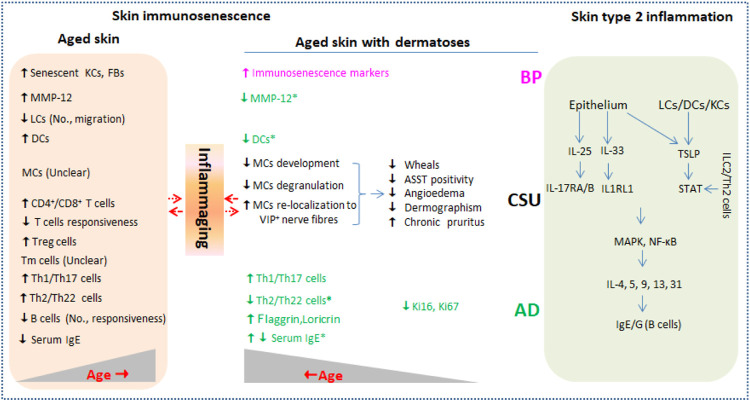
FIGURE 1.
Current evidence of immunosenescence in aged skin with or without type 2 inflammation dermatoses. Type 2 inflammation dermatoses such as AD, CSU, and BP are driven by key cytokines including IL-25, IL-33, and TSLP released from damaged epithelium and LCs/DCs (right column). Skin barrier inevitably undergoes characteristically immunological changes (skin immunosenescence) during aging in healthy individuals (left column) and in individuals affected by type 2 inflammation dermatoses. Inflammaging that is characterized by low level of pro-inflammatory cytokines including CRP, IL-1β, IL-6, and TNF-α produced by senescent skin cells may be in complex interaction with the two conditions. No., number; KCs, keratinocytes; FBs, fibroblasts; LCs, Langerhans cells; DCs, dendritic cells; MCs, mast cells; ILC2, innate lymphoid cell 2; Tm, memory T cells; Ig, immune globulin; MMP-12, matrix metalloproteinase 12; ASST, autologous serum skin test; TSLP, thymic stromal lymphopoietin; STAT, signal transducer and activator of transcription; IL1RL1, IL-1 receptor-like 1; AD, atopic dermatitis; CSU, chronic spontaneous urticaria; BP, bullous pemphigoid.

