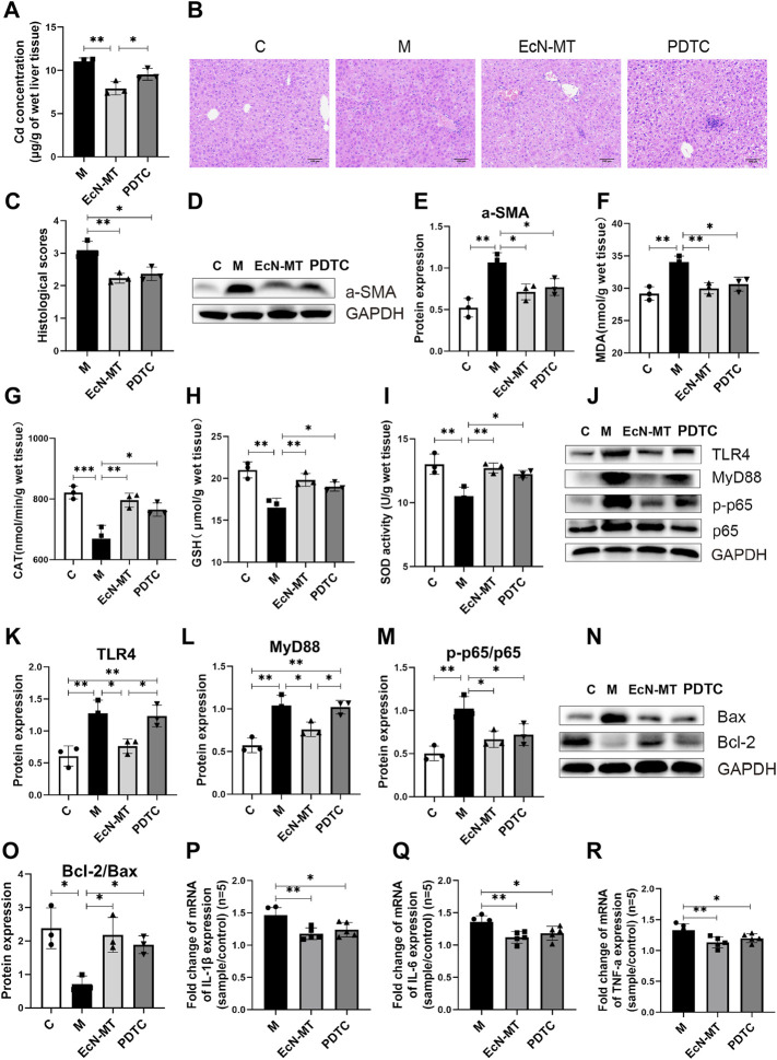
FIGURE 5.
Suppression of NF-κB activation reduced hepatic inflammation and oxidative stress. Values are presented as means ± SD (n = 3, except q-PCR results repeated five times). (A) Cd concentrations in the liver assayed by ICP-MS. (B) HE staining image of liver tissue (200×). (C) Histological score of liver injury. After 8 weeks of exposure to drinking water, the pathological changes of liver tissues stimulated by cadmium chloride (0.545 mM) were determined. (D) Western bloting analysis of a-SMA expression in liver tissues. (E) The relative expressions of a-SMA were quantified by ImageJ, expressions of a-SMA were quantified by ImageJ. GAPDH was used as an internal control. The activity of MDA; CAT; GSH; SOD, activity of (F) MDA; (G) CAT; (H) GSH; (I) SOD. (J) Western bloting analysis of TLR4, MyD88, p-p65, p65 expression in liver tissues. The relative expressions of (K) TLR4, (L) MyD88, (M) p-p65/p65 were quantified by ImageJ. GAPDH was used as an internal control. (N) Western bloting analysis of Bax, Bcl-2 expression in liver tissues. (O) The relative expressions of Bcl-2/Bax were quantified by ImageJ. GAPDH was used as an internal control. The relative mRNA expressions of (P) IL-1β, (Q) IL-6, (R) TNF-α in liver tissues were detected by q-PCR. *p < 0.05, **p < 0.01, ***p < 0.001.

