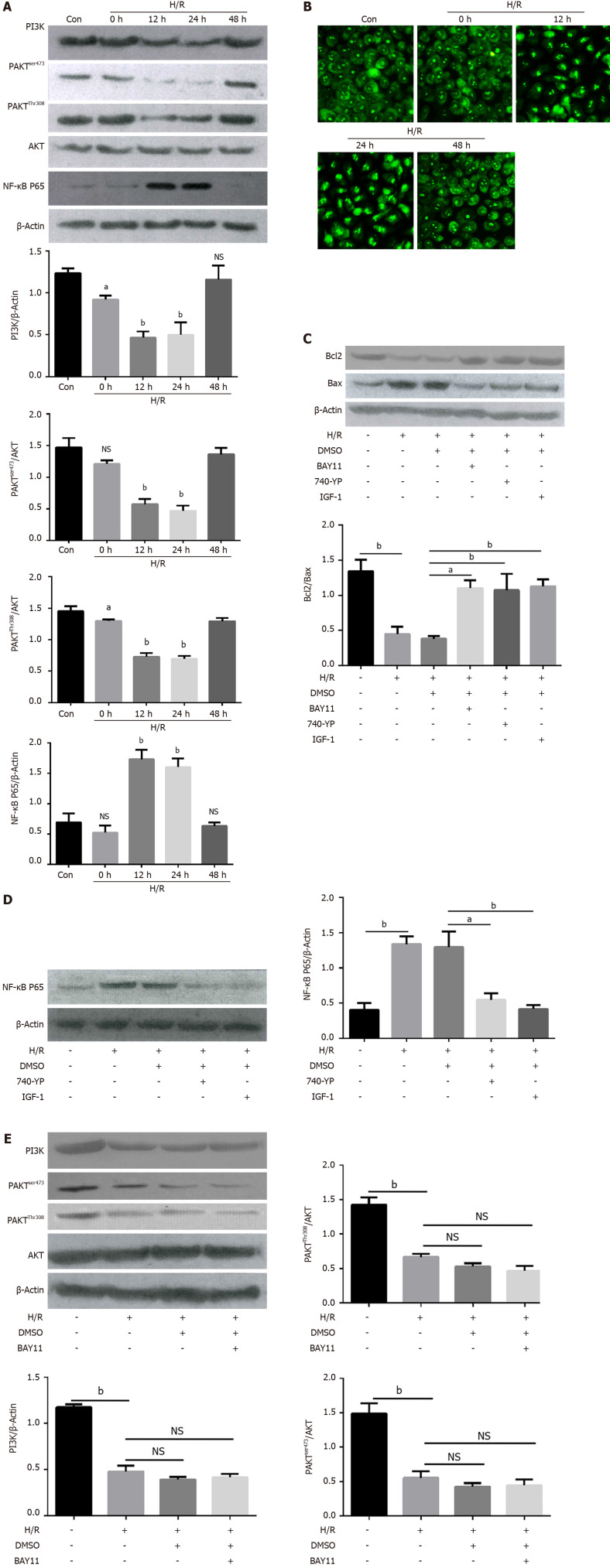
Figure 3.
PI3K/AKT/NF-κB pathway was involved in hypoxia/reoxygenation-induced apoptosis. A: Representative western blotting and quantification data for PI3K, p-AKTser473, p-AKTThr308, NF-κB P65, and β-actin at different time points after hypoxia/reoxygenation (H/R) treatment. ns, no significant difference, aP < 0.01 and bP < 0.001 vs control (n = 3); B: Representative images of immunofluorescence staining for NF-κB P65 in Caco2 cells before (Con) and after the H/R treatment at different time, Bars = 100-μm (n = 3); C: Representative Western blotting and quantification data for cell apoptotic proteins Bcl-2, and Bax after NF-κB inhibitor BAY11, AKT activator IGF-1 and PI3K activator 740 Y-P treatment, aP < 0.01 and bP < 0.001 vs control (n = 3); D: Representative Western blotting and quantification data for NF-κB P65 after AKT activator IGF-1 and PI3K activator 740 Y-P treatment, aP < 0.01 and bP < 0.001 vs control (n = 3); E: Representative Western blotting and quantification data for PI3K, p-AKTser473, and p-AKTThr308, after NF-κB inhibitor BAY11 treatment. Data are presented as mean ± SD. bP < 0.001 vs control (n = 3). NS: No significant difference.

