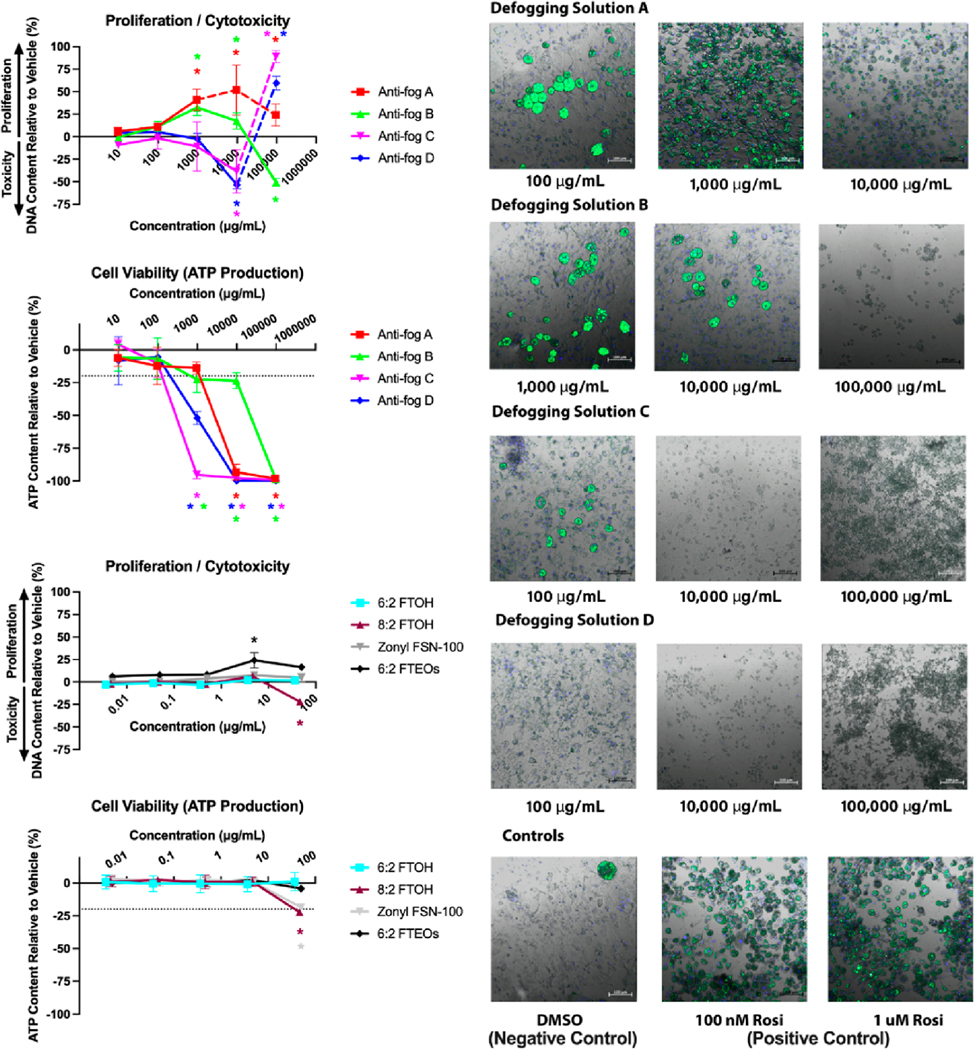
Figure 2.
Cytotoxicity and cell health measures for anti-fog sprays and constituent chemicals. 3T3-L1 pre-adipocytes were differentiated while exposed to sprays and constituent chemicals and then assayed for DNA content (cytotoxicity), ATP production (cell viability), and fluorescent microscopy (qualitative visual confirmation). The DNA content reported as increase (pre-adipocyte proliferation) or decrease (cytotoxicity) relative to differentiated solvent control response. ATP production reported as a decrease in ATP produced relative to differentiated solvent control response. Data presented as mean ± SEM from three independent experiments. Fluorescence microscopy used as a third confirmatory measure of toxicity for anti-fog sprays (green fluorescence measures triglyceride accumulation staining and blue fluorescence represents nuclear staining).

