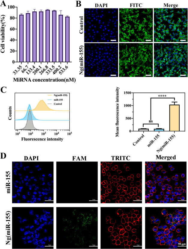
Figure 3.
(A) RAW264.7 cell viability in different concentration gradients of Ng(miR-155). (B) NIH 3T3 cell morphology after being treated by Ng(miR-155). Cells were counterstained with DAPI (nuclei) and FITC-labeled phalloidin (actin). (C) Flow cytometric analyses of RAW264.7 cells after incubation with free miR-155 or Ng(miR-155). Quantitative analyses of fluorescence were shown by mean fluorescence intensity. (D) Confocal laser scanning microscopy images of the RAW264.7 cells incubated with free miR-155 and Ng(miR-155). Cells were counterstained with DAPI (nuclei) and TRITC-labeled phalloidin (actin). Scale bars are 50 μm. ***P < 0.001, ns means no significant difference.

