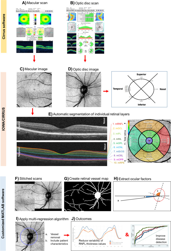
Fig. 1.
Steps to account for ocular factors from circumpapillary retinal nerve fiber layer (cpRNFL) measurement. A, B Capture the optical coherence tomography (OCT) scan protocols using Cirrus (Zeiss) system, one centered in the macula and the other centered in the optic disc. C–E Extract the cpRNFL measurements using Cirrus Review software and the individual macular layers using Iowa Reference Algorithms version 3.8.0 of the OCT layer segmentation program. F Register and stitch the macular and optic disc images. G Segment the retinal vessels to obtain the vessel tree. H Extract the optic disc and fovea features. I Calculate the cpRNFL retinal thickness, using a multi-regression compensation model. J Finally, the ideal model would reduce the variability of cpRNFL thickness measurements and/or improve disease detection

