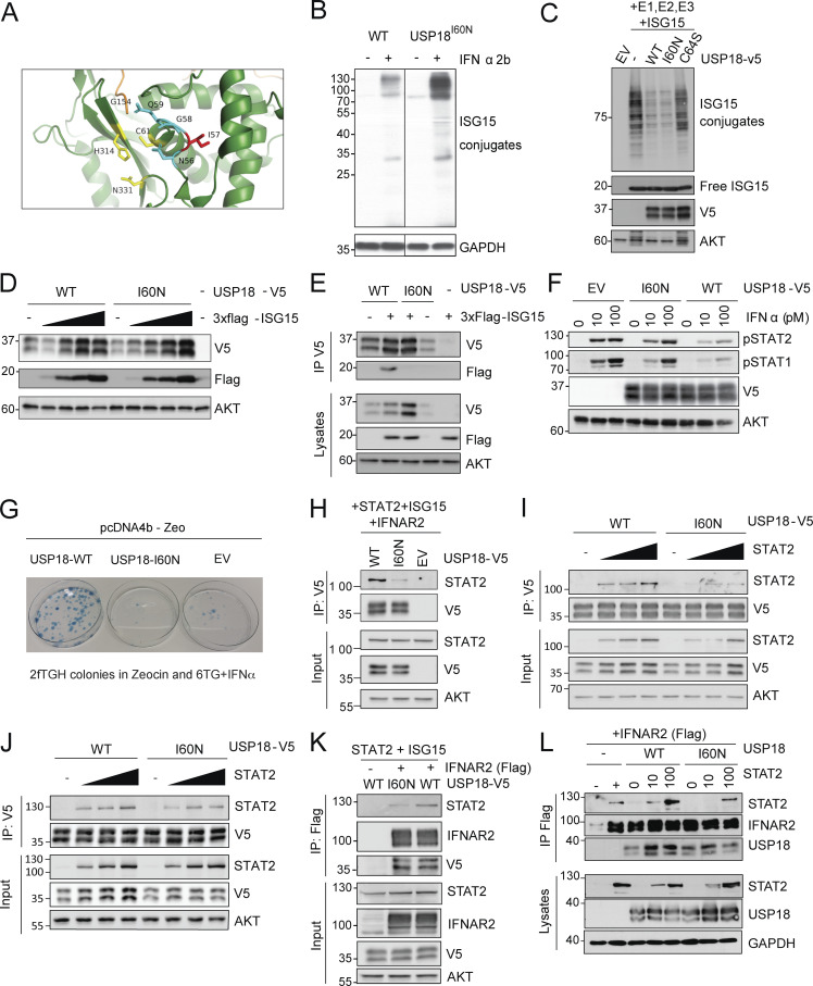
Figure 2.
Characterization of the enzymatic and regulatory functions of USP18 allele. (A) Close-up view of the catalytic cleft of mUsp18 (green, chain A) complexed to mIsg15 (orange; Basters et al., 2017; PDB entry: 5CHV). Residues of the catalytic triad (C61, H314, and N331) are shown in yellow. I57 (I60 in hUSP18) is located in a disordered loop above the catalytic triad. I57 is shown in red, and the neighboring residues of the loop (N56, G58, and Q59) in cyan. Of note, the side chain of I57 points toward an α-helix (aa 117–126), while the side chains of the neighboring N56, G58, and Q59 residues orient toward the catalytic triad. Figure made using PyMOL. (B) hTERT-immortalized fibroblasts from a control donor (WT) or P1 (USP18I60N) were treated with 1,000 IU/ml IFN-α2b for 24 h. Cell lysates were analyzed by Western blot with the indicated antibodies; representative experiment shown. (C) 293T cells were cotransfected with UbE1L (E1), UbCH8 (E2), HerC5 (E3), and 3xFlag-ISG15 in combination with USP18-V5 (WT), the c.179T>A variant (I60N), or the catalytically inactive mutant C64S. Cell lysates were analyzed by Western blot as indicated. ISG15 (conjugates and free) was detected with anti-ISG15 antibodies (a gift of E.J. Borden). In this and all panels below, USP18 was detected using V5 antibodies. (D) 293T cells were cotransfected with USP18-V5 WT or I60N and increasing amounts of 3xFlag-ISG15. Cell lysates were analyzed by Western blot as indicated. (E) 293T cells were cotransfected with USP18-V5 WT or I60N with 3xFlag-ISG15, as indicated. Immunoprecipitation (IP) of USP18 was performed using V5 antibodies. Immunoprecipitates were analyzed by Western blot. (F) 293T cells were transfected with EV, USP18-V5, WT, or I60N. Cells were then treated with the indicated doses of IFN-α2 for 20 min. Fresh lysates were analyzed by Western blot as indicated. (G) 2fTGH cells were transfected with empty pcDNA4b vector (EV), USP18-WT, or -I60N. Transfected cells were seeded in Zeocin-containing medium. 11 d later, fresh medium with Zeocin and 6TG plus IFN-α2b was added. Colonies were fixed and stained 7 d later. Only cells not responding to IFN, i.e., expressing functional USP18, survive. (H) 293T cells were cotransfected with USP18-V5, WT, or I60N or EV together with STAT2, IFNAR2-Flag, and ISG15. Immunoprecipitation of USP18 was performed using V5 antibodies. (I) 293T cells (p60 dish) were cotransfected with 650 ng of USP18-V5 WT or I60N and increasing amounts of STAT2 (210, 420, and 650 ng). 24 h after transfection, USP18 was immunoprecipitated using V5 antibodies. Immunoprecipitates were analyzed by Western blot with STAT2 and V5 antibodies. (J) Experiment as described in I, except that cells were harvested 48 h after transfection. In the immunoprecipitate, USP18 was revealed using USP18 antibodies. (K) 293T cells were cotransfected with USP18-V5, WT, or I60N together with STAT2, IFNAR2-Flag, and ISG15. Immunoprecipitation of IFNAR2 was performed using Flag antibodies. (L) 293T cells were cotransfected with USP18-V5, WT, or I60N together with IFNAR2-Flag and increasing amounts of STAT2 (10 and 100 ng). Immunoprecipitation of IFNAR2 (∼90–100 kD) was performed using Flag antibodies. All results in the figure are representative of at least two independent experiments. The molecular weight markers (kD) are shown on the left.

