Figure 3.
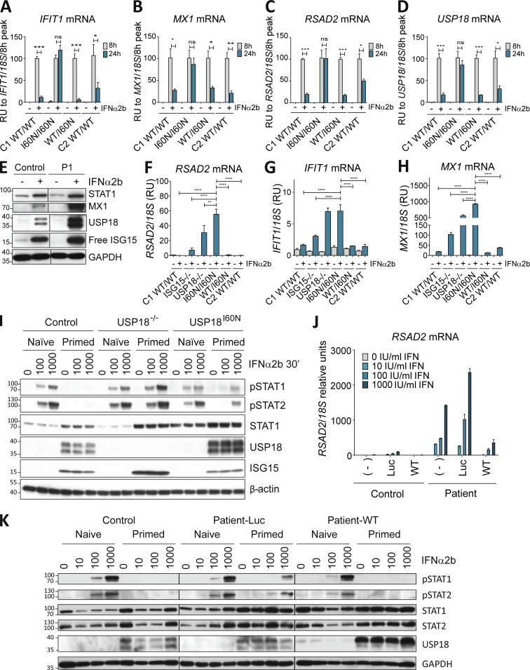
Accumulation of USP18 I60N protects from subsequent IFN challenges. (A–D) hTERT-immortalized fibroblasts from control donor (C1 WT/WT), P1 (I60N/I60N), heterozygous parent (WT/I60N), or healthy brother (C2 WT/WT) were treated with 1,000 IU/ml IFN-α2b for 8 or 24 h. Relative mRNA levels were assessed for IFIT1, MX1, RSAD2, and USP18 performed three times each with technical triplicates. The results are represented relative to 8 h as the peak of ISG induction. A representative experiment is shown. Data analysis was performed with unpaired t tests. ns, P > 0.05; *, P < 0.01; **, P < 0.001; ***, P < 0.0001. (E) hTert-immortalized fibroblasts from a control donor (Control) or P1 were treated with 1,000 IU/ml IFN-α2b for 24 h. Cell lysates were analyzed by Western blot for the indicated antibodies; representative experiment shown. (F–H) hTERT-immortalized fibroblasts from a control donor (C1 WT/WT), ISG15-deficient donor (ISG15−/−), USP18-deficient donor (USP18−/−), P1 (I60N/I60N), heterozygous parent (WT/I60N), or healthy brother (C2 WT/WT) treated with 1,000 IU/ml IFN-α2b for 12 h, washed with PBS, and left to rest for 36 h. Relative mRNA levels were assessed for RSAD2, IFIT1, and MX1, performed three times each with technical triplicates; representative experiment shown. Bars represent the mean ± SEM. Statistical analysis performed by one-way ANOVA. **, P < 0.001; ****, P <0.00001. (I) hTERT-immortalized fibroblasts from control donor (Control), USP8-deficient donor (USP18−/−), and P1 (USP18I60N) were primed with 1,000 IU/ml IFN-α2b for 12 h, washed, left to rest for 36 h, and restimulated with increasing amounts of IFN-α2b for 30 min. Cell lysates were analyzed by Western blot for the indicated antibodies; representative experiment shown. (J) hTert-immortalized fibroblasts from a control donor (Control) or P1 were mock-transduced (−) or transduced with Luc-RFP (Luc) or WT USP18 (WT), sorted, treated with the indicated doses of IFN-α2b for 12 h, washed with PBS, and left to rest for 36 h, after which relative mRNA levels were assessed for RSAD2, performed three times each with technical triplicates; representative experiment shown. Bars represent the mean ± SD. (K) hTert-immortalized fibroblasts from control donor (Control), P1 transduced with Luc-RFP (Patient-Luc), or P1 transduced with WT USP18 (Patient-WT) were primed with 1,000 IU/ml IFN-α2b for 12 h, washed, left to rest for 36 h, and restimulated with IFN-α2b for 20 min. Cell lysates were analyzed by Western blot with the indicated antibodies. All results in the figure are representative of at least two independent experiments. The molecular weight markers (kD) are shown on the left. RU, relative units.

