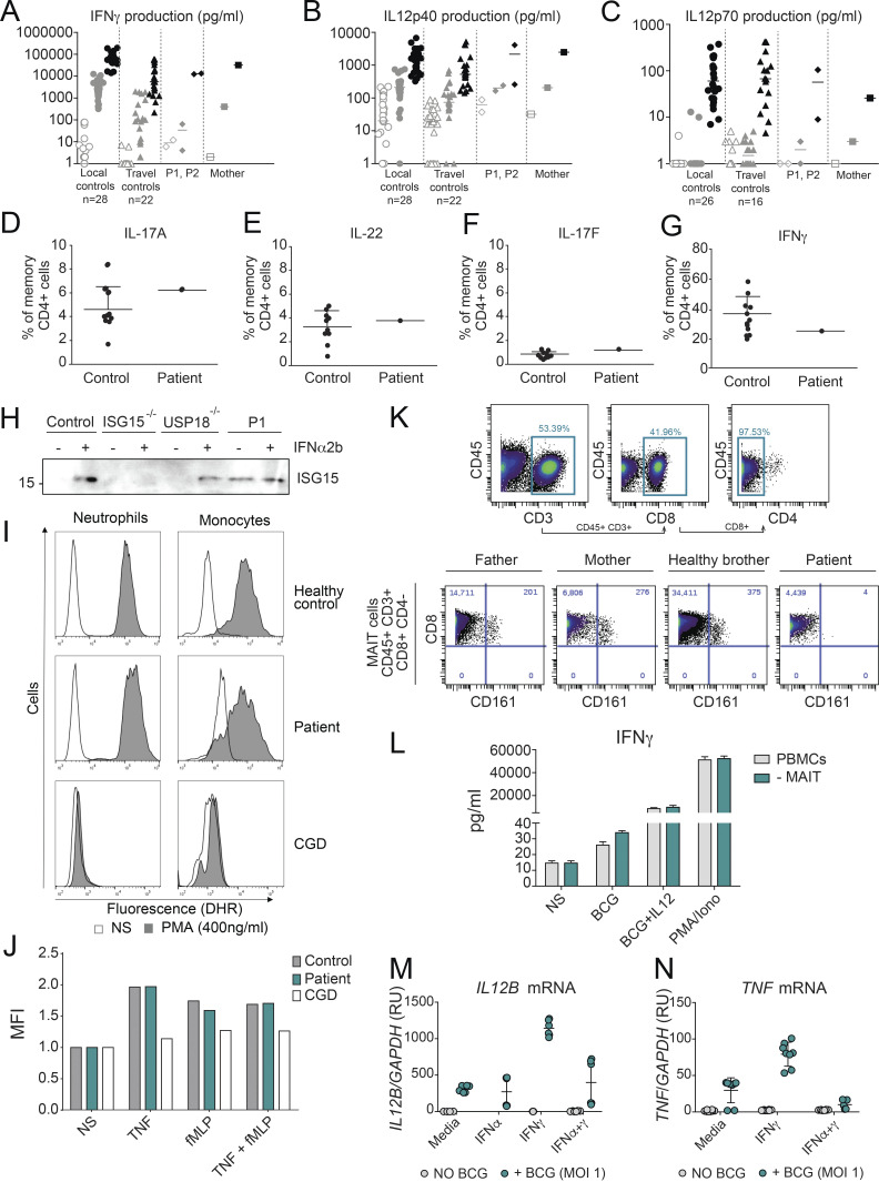
Figure S4.
Characterization of the MSMD phenotype caused by the USP18 I60N variant. (A–C) Cytokine production in the supernatants of whole-blood cells from local controls (n = 28), travel controls (n = 22), P1, P2, and mother, left unstimulated or stimulated with BCG alone or BCG plus cytokine (indicated), as detected by ELISA. (D–G) The ability to produce IL-22, IL-17A, IL-17F, and IFNγ by CD4+ T cells was assayed in Control and USP18 I60N patient. (H) Control and P1 fibroblasts were stimulated with 1,000 U/ml of IFN-α2b for 24 h. The presence of secreted ISG15 was analyzed by immunoblotting of the supernatant proteins. The molecular weight markers (kD) are shown on the left.(I) Dihydrorhodamine 123 (DHR) fluorescence was assayed using samples from healthy control and I60N patient neutrophils and monocytes treated with or without PMA. (J) DHR fluorescence was assayed using samples from healthy control, I60N patient, and CGD patient neutrophils treated with the indicated stimuli. MFI, mean fluorescence intensity. (K) Gating strategy for the quantification and identification of MAIT cells in PBMCs from father, mother, healthy brother, and P1 analyzed by CyTOF. (L) IFN-γ production was measured by ELISA in the supernatants of total PBMCs and MAIT-depleted PBMCs that were left unstimulated, stimulated with BCG alone or BCG plus IL-12, or stimulated with PMA/ionomycin. (M and N) Control monocytes were treated with 1,000 IU/ml IFN-α2b and 1,000 U/ml IFN-γ overnight, washed, and stimulated with BCG for 18 h, followed by measurement of mRNA for IL12B and TNF. All results are representative of at least two independent experiments. MOI, multiplicity of infection; RU, relative units.

