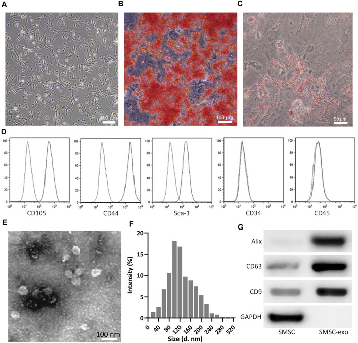
FIGURE 2.
Isolation of SMSCs and SMSC-derived exosomes (SMSC-Exos). (A) Observation of SMSCs morphology at passage 3. (B) After 14 days of osteogenic differentiation, matrix mineralization of SMSCs was stained with Alizarin Red S. (C) after 14 days of adipogenesis induction, SMSCs were stained with Oil Red O. (D) Fluorescence Activated Cell Sorting was performed on SMSCs for CD105, CD44, Sca-1, CD34, and CD45 at passage 3. (E) Identification of exosomes by TEM. (F) Nanoparticle Tracking Analysis of the isolated exosome. (G) Detection of CD9, CD63, and Alix expression by Western blot analysis between SMSCs and SMSC-derived exosomes (SMSC-Exos).

