Figure 2.
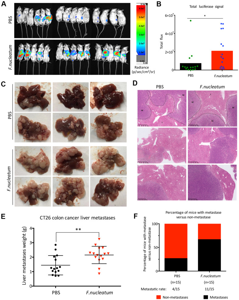
Administration of Fusobacterium nucleatum promotes liver metastasis of colorectal cancer in mice. (A, B) PBS-resuspended 109 colony-forming units of F. nucleatum or PBS were administrated to mice for 8 weeks by gavage every day. The mice received intra-splenic injection post 2-week administration of F. nucleatum or PBS intragastrically. At day 28 after intra-splenic transplantation of CT26-Luc cells, in vivo bioluminescence imaging was used to assess liver metastases (A) of BALB/c mice (n=15 per group). (B) The corresponding data expressed by the change in the total signal flux. Data are presented as mean ± standard deviation (SD; *P < 0.05, **P < 0.01, and ***P < 0.001; unpaired Student’s t-test). (C, D) After the 2-week treatment of F. nucleatum or PBS, mice received intra-splenic injection of CT26 tumor cells. Four weeks later, the mice were euthanized, and subsequent histological studies were performed. The representative metastatic foci (C) and hematoxylin and eosin-stained liver sections (D) in mice are shown. (E, F) Livers were weighed (E) and liver metastatic rates were measured (F). Data are presented as mean ± SD (*P < 0.05, **P < 0.01, and ***P < 0.001; unpaired Student’s t-test).

