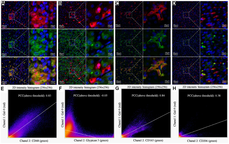
Figure 4.
Representative dual IF staining result to show the colocalization of Gal-9 with different cell markers. Dual-IF staining of Gal-9 and CD68 (A), Gal-9 and GPC3 (B), Gal-9 and CD163 (C), Gal-9 and CD206 (D). (blue: nuclei; red: Gal-9; green: CD68/GPC3/CD163/CD206; yellow: merge). Quantitation of the colocalization of Gal-9 and CD68/GPC3/CD163/CD206 is shown in (E–H). PCCs (above threshold) of Gal-9 and CD68/GPC3/CD163/CD206 were 0.83, -0.05, 0.84 and 0.38, respectively. Results showed that Gal-9 was mostly expressed on CD68+CD163+ KCs, but not HCC tumor cells or M2 macrophages. Scale bar, 50μm (left column) and 10μm (right column).

