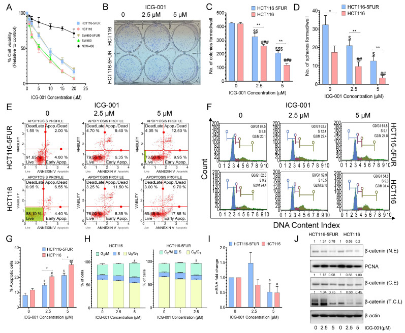
Figure 3.
Anti-proliferative effects of the Wnt signaling inhibitor ICG-001 in parental and 5FUR CRC cells. (A) Measurement of percent cell viability after treatment with ICG-001 (1–20 mM) for 48 h in parental and 5FUR CRC cells by CCK-8 assay. (B) Representative images of colonies and (C) the number of colonies formed after treatment with different concentrations of ICG-001 for 48 h. (D) Sphere-formation capacity after treatment with ICG-001 for 48 h. (E). Representative histogram of the percentage of apoptotic cells and (G) a graphical representation of the percentage of apoptotic cells after treatment with different concentrations of ICG-001 in parental and HCT116-5FUR cells for 48 h. (F) Representative histograms of different phases of the cell cycle and (H) a graphical representation of the percentage of cells in different phases of the cell cycle after treatment with different concentrations of ICG-001 for 48 h. (I) Gene and (J) protein expression analyses of β-catenin in parental and HCT116-5FUR cells after treatment with ICG-001 for 48 h (N.E-nuclear extract, C.E-cytoplasmic extract, T.C.L-total cell lysates). Original blots see Figure S2. Statistical significance was determined by a Student’s t-test. (Comparison between Parental vs. 5FU-resistant group-* p < 0.05, ** p < 0.01; comparison between Control and treatment groups for HCT116-$ p < 0.05, $$ p < 0.01, $$$ p < 0.001; comparison between Control and treatment groups for HCT116-5FUR-# p < 0.05, ## p < 0.01, ### p < 0.001.)

