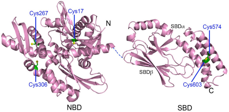
Figure 2.
Crystal structures of human HspA1A. The nucleotide-binding domain (NBD) in the ADP-bound state (PDB code 3AY9) and the substrate-binding domain (SBD, PDB code 4PO2) are shown. The dashed line represents the flexible linker between the NBD and SBD. The SBD contains a β-sandwich substrate-binding subdomain (SBDβ) and an α-helical lid subdomain (SBDα). The five Cys residues are labeled in green. Figure reproduced from ref. [52].

