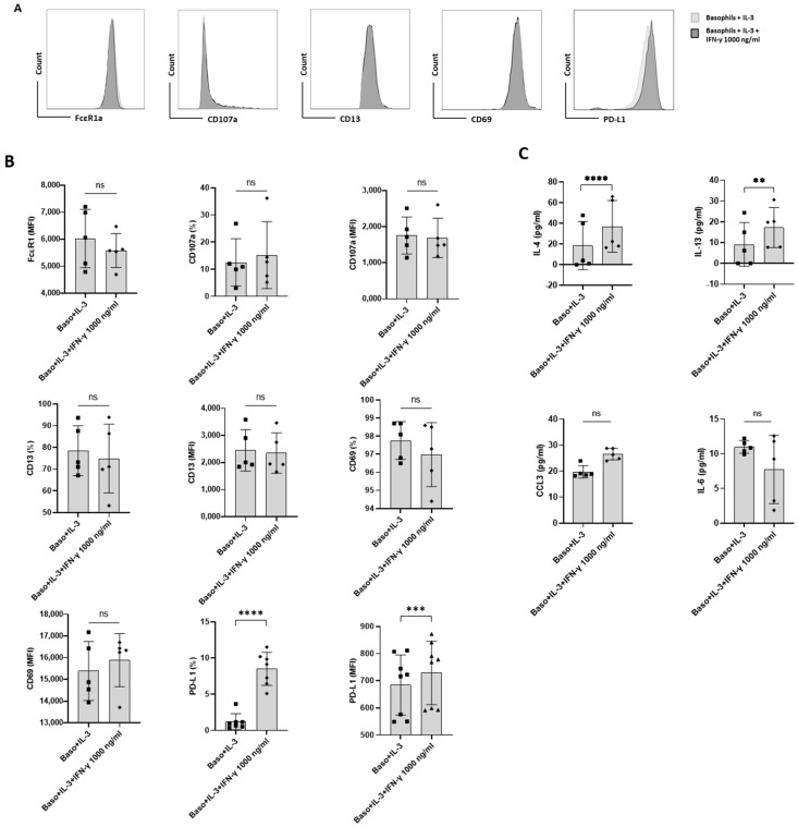
Figure 2.
The effect of IFN-γ on PD-L1 expression in primed primary human basophils. Basophils (0.1 × 10⁶ cells/200 μL/96-well plate) isolated from PBMCs of healthy donors were cultured with or without IFN-γ at 1000 ng/mL. Simultaneously, basophils were primed with IL-3 along with IFN-γ treatment. Basophil phenotype was evaluated by flow cytometry after 24 h. (A) Gating strategy and representative histogram overlays displaying the expression pattern of FcεR1, CD107a, CD13, CD69, and PD-L1. (B) Expression of FcεR1, CD107a, CD13, CD69, and PD-L1 on basophils (% positive cells and median fluorescence intensities (MFI), mean ± SD; n = 5–8 independent donors with three independent experiments). (C) The amount (pg/mL) of secreted IL-4, IL-13, CCL3 and IL-6 in the cell-free supernatant from the above experiments. The data were presented as the mean ± SD and were from 5–8 independent donors and three independent experiments). ns, not significant, ** p < 0.01, *** p < 0.001, **** p < 0.0001, paired Wilcoxon test.

