Figure 3.
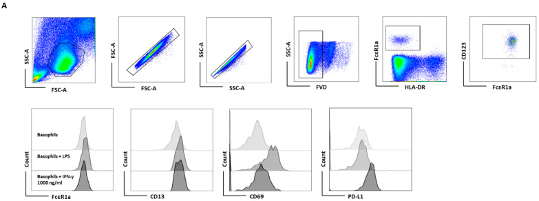
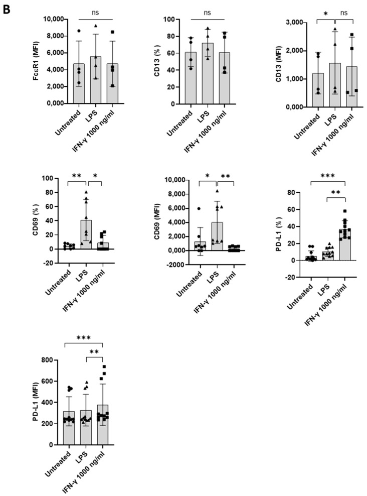
IFN-γ-induced PD-L1 expression in basophil is legible in PBMCs. Basophil-containing PBMCs (1 × 10⁶ cells/mL/24-well plate) from the healthy donors were cultured with or without IFN-γ at 1000 ng/mL or LPS at 100 ng/mL for 24 h. After incubation, cells’ phenotype was evaluated by flow cytometry. (A) Gating strategy and representative histogram overlays are displaying the expression pattern of FcεR1, CD107a, CD13, CD69, and PD-L1 on the basophils. (B) Expression of FcεR1, CD13, CD69, and PD-L1 on the basophils (% positive cells and median fluorescence intensities (MFI), mean ± SD; n = 4–11 independent donors from three independent experiments). ns, not significant, * p < 0.05, ** p < 0.01, *** p < 0.001, one-way ANOVA Friedman test with Dunn’s multiple comparisons post-test.


