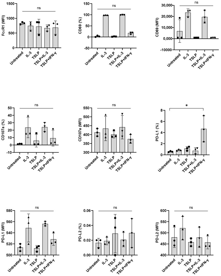
Figure 5.
The effect of TSLP on the expression of PD-L1 induced by IFN-γ in primary human basophils. Basophils (0.1 × 10⁶ cells/200 μL/96-well plate) isolated from PBMCs of healthy donors were cultured with either IL-3, TSLP (10 ng/mL), TSLP + IL-3 or TSLP + IFN-γ. Basophil phenotype was evaluated by flow cytometry after 24 h. Expression of FcεRI, CD69, CD107a, PD-L1, and PD-L2 on the basophils (% positive cells and median fluorescence intensities (MFI), mean ± SD; n = 3 independent donors from three independent experiments) was presented. ns, not significant, * p < 0.05, one-way ANOVA Friedman test with Dunn’s multiple comparisons post-test.

