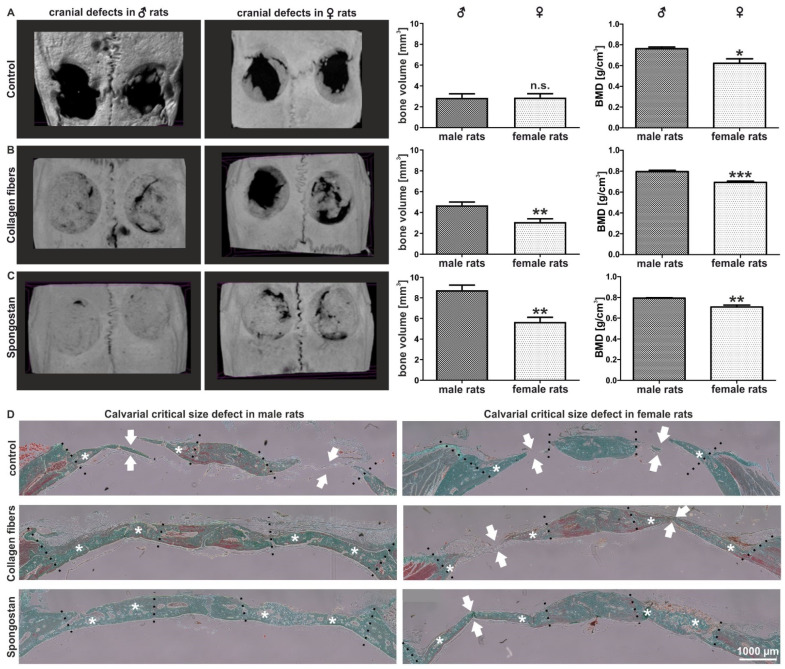
Figure 1.
Female rats showed a reduced capacity for regenerating calvarial bone lesions after transplantation of Spongostan or collagen type I fibers into critical-size calvarial defects. (A) Micro-computed tomography (µCT) followed by quantification of bone volume and bone mineral density (BMD) revealed no closure of the critical-size defects in female or control male animals, but a significantly decreased BMD in female control rats. Mann–Whitney test, * p < 0.05, ** p < 0.01, *** p < 0.001 was considered significant. (B,C) µCT scans depicting a significantly decreased bone volume and BMD in female animals compared to their male counterparts after transplantation of collagen fibers or Spongostan. (D) Histological examination revealed elevated levels of newly formed bone in male animals upon transplantation of collagen fibers or Spongostan in male rats. Stars mark newly formed bone; arrows indicate unclosed lesions.

