Figure 2.
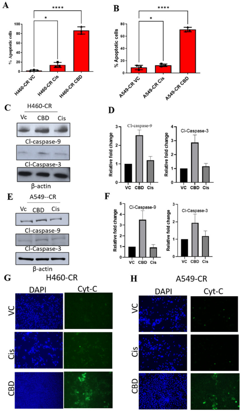
CBD-mediated induction of apoptosis in CR NSCLC cells. (A) H460-CR or (B) A549-CR cells were treated with VC, cisplatin, or CBD. Percentage apoptotic cells were analyzed using Annexin-V and 7-AAD staining and analyzed by flow cytometry analysis. (C,E) H460-CR or A549-CR cells treated with VC or CBD or Cisplatin and cell lysates were evaluated for caspase 3 and caspase 9 apoptotic markers by Western blot analysis. β-actin was used as loading control. (D,F) Graphs represents densitometric ratio in fold change for H460-CR or A549-CR respectively. (G), H460-CR or (H) A549-CR cells were treated with VC, cisplatin, or CBD, and cytochrome-c release was measured by immunofluorescence, all the images were taken at 10X magnification. Data are represented as Mean S.D (* p < 0.01, **** p < 0.0001 using one-way ANOVA).

