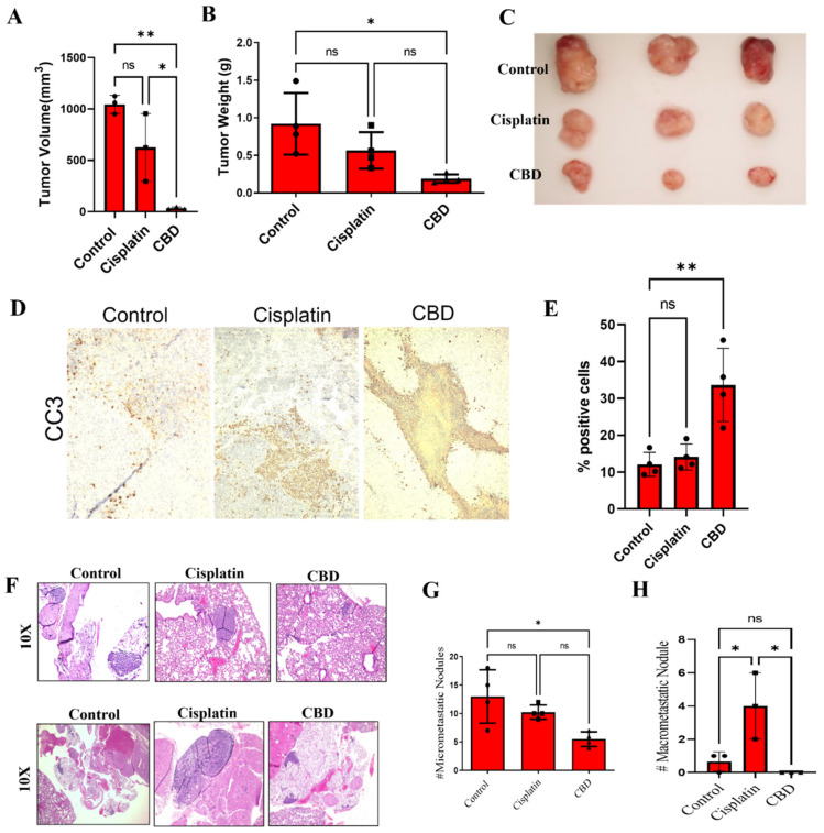
Figure 5.
CBD inhibits CR NSCLC growth and metastasis in vivo. (A–C) H460-CR cells were injected subcutaneously into the right flank of nude mice (n = 5). Palpable tumors were treated with control (PBS) or CBD (10mg/kg body weight) or cisplatin (5 mg/kg body weight) for 4 weeks. (A) The tumor volume was measured externally at the end. (B) Weight of harvested tumors, and (C) the tumors were harvested, and representative images are presented. (D) The tumor sections were processed and immunostained for cleaved-caspase-3 (CC3) and images were taken at 10X magnification. (E) The CC3 staining images from (D) were quantified for CC3 % positive cells. (F–H) The lungs harvested from (A) were processed and H&E stained. (F) Representative H&E images of the lungs were harvested at 10x magnification. Upper and lower panel represents micro- and macro-metastasis, respectively. (G) Quantification of micro- and (H) macro-metastasis using ImageJ analysis. * is p-value < 0.01, ** p < 0.001, and ns is non-significant using one-way Anova.

