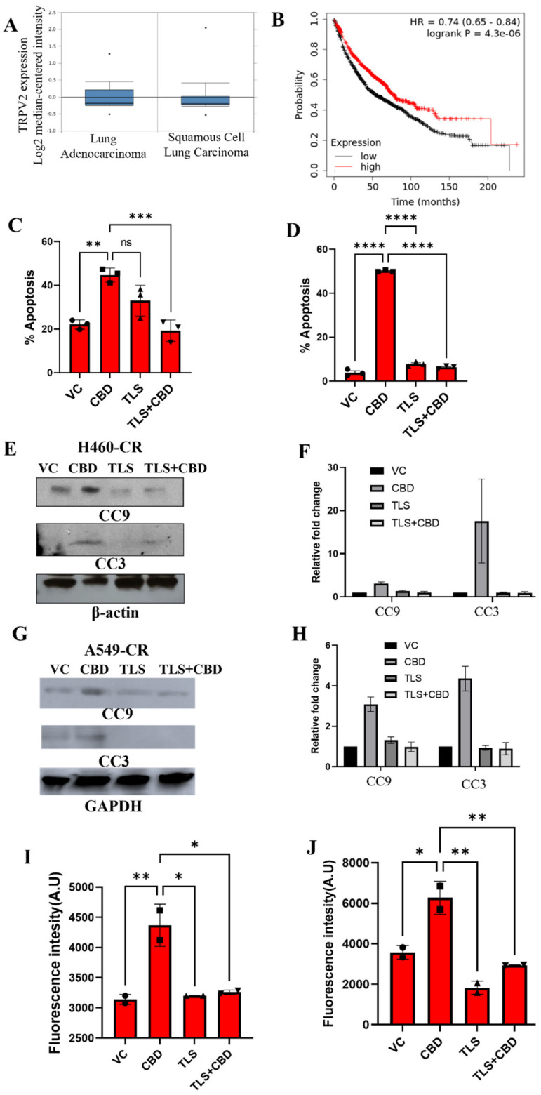
Figure 6.
CBD interacts with TRPV2 channel to induce apoptosis. (A) Analysis of TRPV2 expression in lung adenocarcinoma (n = 63) and squamous cell carcinoma (n = 75) patient tumors using TCGA dataset. (B) The association between TRPV2 expression (low vs high) and overall survival of lung cancer patients was analyzed using KM plotter. (C) H460-CR and (D) A549-CR cell lines were treated with VC or CBD in the presence or absence of TLS. The percentage apoptosis was evaluated using annexin/7-AAD staining and flow cytometry. Cell lysates from (E) H460-CR and (G) A549-CR cell lines treated with VC or CBD in the presence or absence of TLS were analyzed for the expression of apoptosis markers, cleaved caspase 9 (CC9), and cleaved caspase 3 (CC3) by Western blot analysis using beta-actin and GAPDH as loading control. (F,H) Graphs represents densitometric ratio in fold change for (E,G), respectively. (I) H460-CR and (J) A549-CR cell lines were treated with VC or CBD, the presence or absence of TLS, and the free intracellular calcium levels were analyzed by estimating fluorescence intensity of calcium green-1-AM staining. Data are represented as Mean ± S.D (* p < 0.01, ** p < 0.001, *** p < 0.001, **** p < 0.0001, ns; non-significant). Statistical comparisons between two groups were carried out using Students t-test, while a one-way ANOVA was used for more than two groups.

