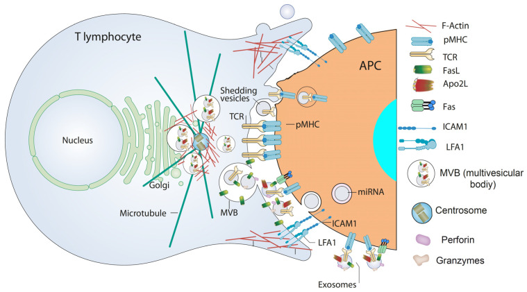
Figure 3.
EV in the immune synapse. In a mature IS produced by TCR stimulation via the peptide-MHC complex (pMHC) on the APC and the interaction of accessory molecules (such as Intercellular Adhesion Molecule 1—ICAM1—with Lymphocyte function-associated antigen 1—LFA-1) F-actin is reduced at the cSMAC, the central region of the IS. F-actin accumulates at the distal SMAC (dSMAC), and F-actin around the centrosome depolymerizes. These F-actin reorganization processes, acting in a coordinated manner, may assist centrosome traffic towards the IS and the simultaneous convergence of MVB towards the F-actin depleted area in the cSMAC, facilitating MVB fusion at the cSMAC, and the subsequent exosome secretion carrying TCR and proapoptotic molecules in the synaptic cleft. In addition, shedding vesicles emerging from the plasma membrane and containing TCR are represented at the synaptic cleft. Both exosomes containing miRNA [15] and shedding vesicles [107,108] are engulfed by APC and provide biological responses in APC. For more details please refer to [12,69,107,109].

