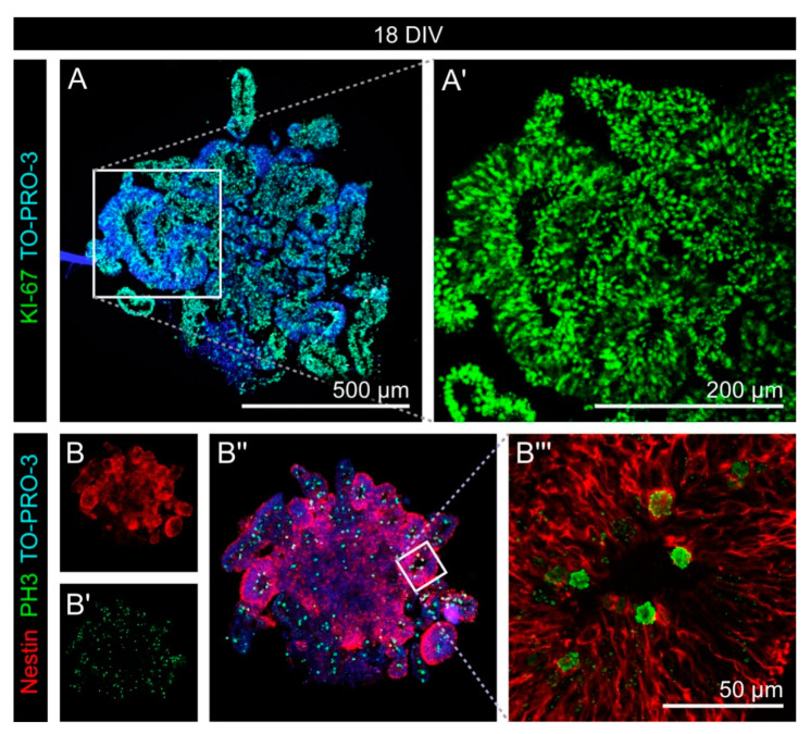
Figure 2.
Proliferation in early cerebral organoids. (A,A′) KI-67 (green), which labels all proliferating cells, was expressed by the vast majority of all cells labeled with nuclear TO-PRO-3 staining (blue). (B–B‴) Phospho-Histone H3 (green), a marker restricted to late G2- and M-phase cells, was expressed by a minority of cells. Within the neural rosette-like structures formed by Nestin-positive cells (red), PH3 signals were primarily found at the apical side, near the lumen.

