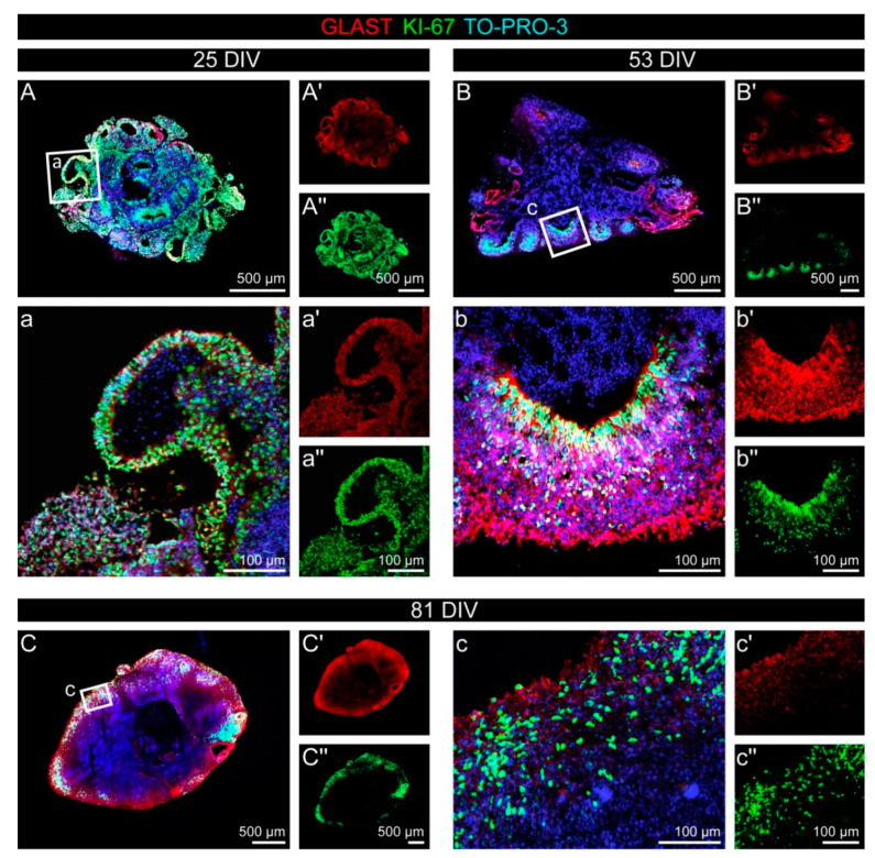
Figure 3.
Proliferation in cerebral organoids over time. (A–c″) The ratio of proliferating cells was reduced over time, as the comparison of KI-67 (green) in an early cerebral organoid after 25 DIV (A–a″) with the situation after 53 DIV (B–b″) or even 81 DIV (C–c″) shows. GLAST (red), a marker for the radial glia type of neural stem cells, was strongly expressed in the neural rosette structures in the early organoids, whereas the signal appeared more diffuse after 81 DIV.

