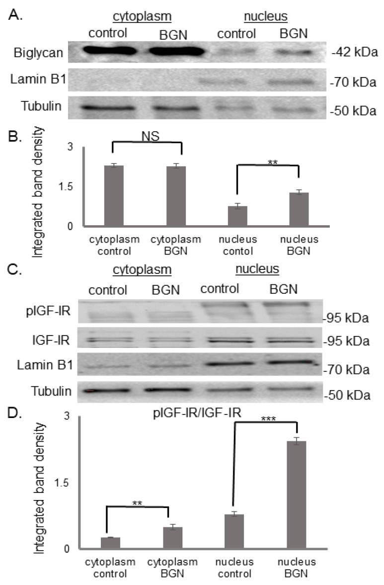
Figure 2.
Effect of biglycan on its own deposition pIGF-IR protein expression at the different MG63 cell compartments. Expression of (A) biglycan and (C) pIGF-IR and in the cytoplasmic compartment of the cells treated with 0% FBS-medium (cytoplasm control) and cells treated with biglycan 10 μg/mL (cytoplasm BGN), as well as the nuclear compartment of the cells (nucleus control; nucleus BGN) were determined by Western blot analysis. Purity controls tubulin and lamin B1 were used for cytoplasmic and nuclear proteins, respectively. Equal amounts of protein from each compartment were loaded, and (B,D) densitometric analysis was performed and plotted. Representative blots are presented. Results represent the average of three separate experiments. Means ± S.E.M were plotted; statistical significance: ** p ≤ 0.01, *** p ≤ 0.001, not significant (NS) compared with the respective control samples.

