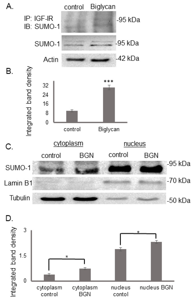
Figure 6.
Effect of biglycan on colocalization of IGF-IR with SUMO-1 and SUMO-1 deposition in the cell. Control cells were treated with serum-free culture medium, and samples were treated for 48 with biglycan (10 μg/mL). (A) Cells extracts were incubated with IGF-IR antibody overnight in a rotating platform and IGF-IR complexes were immunoprecipitated with Protein A/G. Western blot analysis was used for the visualization of SUMO-1 protein immunoprecipitation with IGF-IR. (B) Densitometric analysis of the bands of immunoprecipitated proteins was normalized against the total expression of each protein in the cells and plotted. (C) Expression of SUMO-1 in the cytoplasmic compartment of the cells treated with 0% FBS-medium (cytoplasm control) and cells treated with biglycan 10 μg/mL (cytoplasm BGN), as well as the nuclear compartment of the cells (nucleus control; nucleus BGN) were determined by Western blot analysis. Purity controls tubulin and lamin B1 were used for cytoplasmic and nuclear proteins, respectively. Equal amounts of protein from each compartment were loaded, and (D) densitometric analysis was performed and plotted. Representative blots are presented. Results represent the average of three separate experiments. Means ± S.E.M were plotted; statistical significance: * p ≤ 0.05, *** p ≤ 0.001 compared with the respective control samples.

