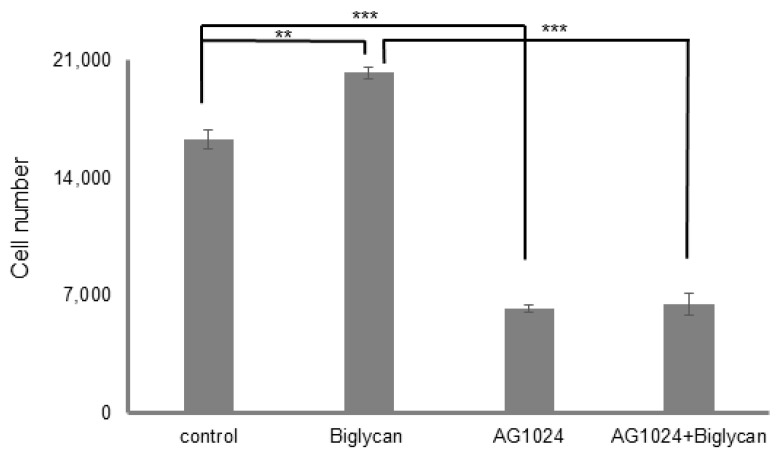
Figure 8.
Role of IGF-IR on biglycan-dependent MG63 cell proliferation. MG63 cells were harvested and seeded on 96-well plates and were allowed to rest overnight. The next day, cells were treated with serum-free medium for 24 h. Cells, in each well, incubated with 0% FBS-medium (control), 10 μg/mL biglycan, 10 µM AG1024 and 10 μg/mL biglycan + 10 µM AG1024 for 48 h (30 min pre-treatment). Cells were counted using fluorometric CyQUANT assay kit. Results represent the average of three separate experiments. Means ± S.E.M were plotted; statistical significance: *** p ≤ 0.001, ** p ≤ 0.01 compared with the respective control samples.

