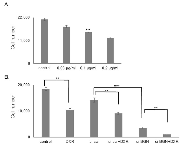
Figure 11.
Effect of biglycan and doxorubicin on MG63 cells proliferation. MG63 cells were harvested and seeded (1500 cells/well) on 96-well plates and transfection with siRNAs (short interfering RNAs) was performed, when needed. (A) Control cells were treated with completed culture medium and samples were treated with different doxorubicin concentrations for 48 h. (B) Cells, in each well, were incubated in serum-free medium and transfected with either siRNAs against biglycan (siBGN) or scrambled siRNAs (siScr), used as negative control. After 24 h, the medium was replaced with 10% FBS DMEM and cells were incubated with 0.1 μg/mL doxorubicin for 48 h. Cells, in each well, were counted after an incubation period, using fluorometric CyQUANT assay kit. Results represent the average of three separate experiments. Means ± S.E.M were plotted; statistical significance: ** p ≤ 0.01, *** p ≤ 0.001 compared with the respective control samples.

