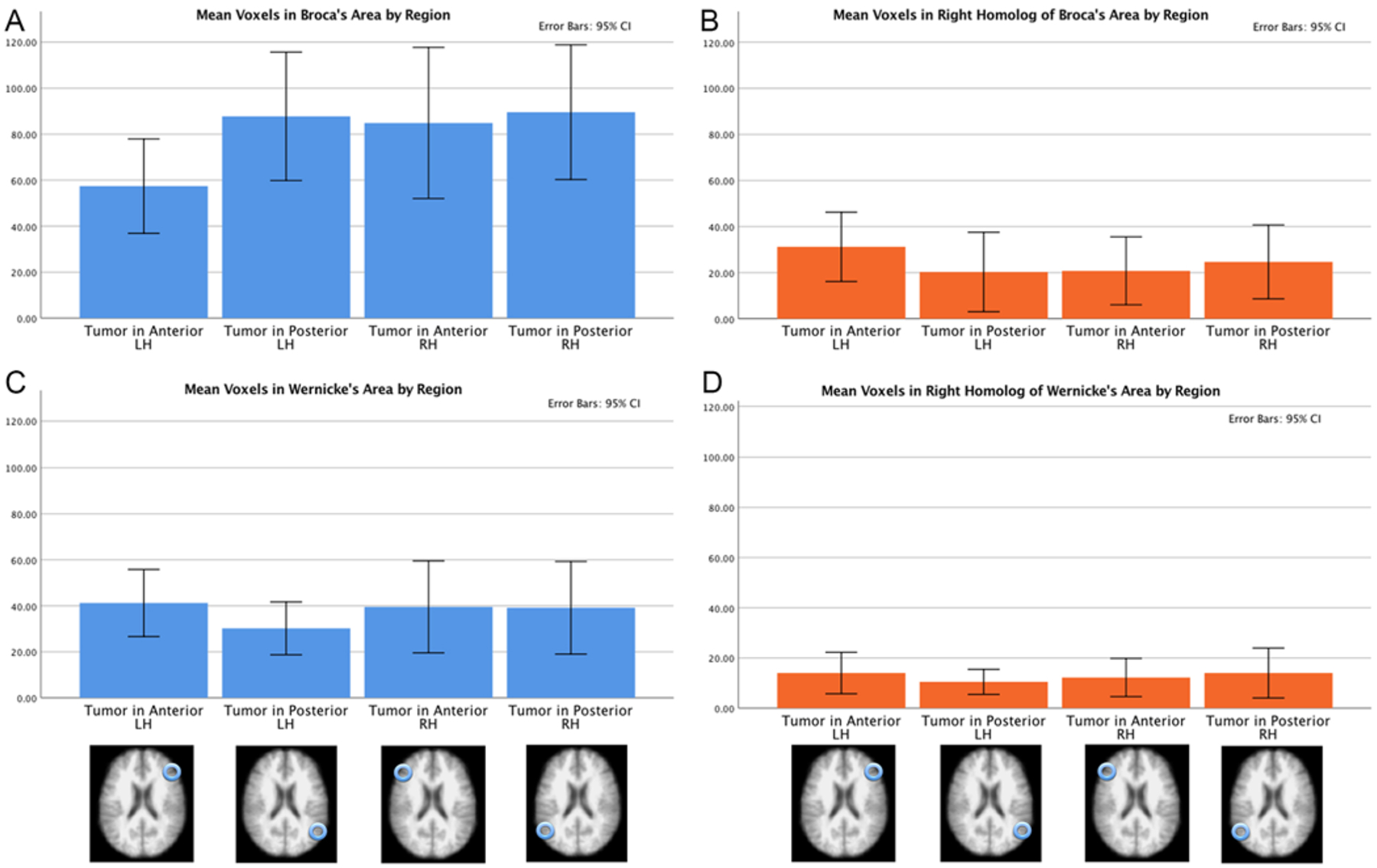
FIG. 5.

Active voxel counts in 3 fMRI language tasks in 4 patient groups by ROI. The results were obtained from 4 patient groups that differed based on their tumor location: the anterior right hemisphere, left anterior hemisphere, posterior right hemisphere, and posterior left hemisphere. The 4 ROIs are Broca’s area (A), the right hemisphere homolog of Broca’s area (B), Wernicke’s area (C), and the right hemisphere homolog of Wernicke’s area (D). In Broca’s area, there was decreased activity in the left anterior group. In the right hemisphere homolog of Broca’s area, there was slightly increased activity in the left anterior group. In Wernicke’s area, there was slightly decreased activity in the left posterior group. The amount of activity in the right hemisphere homolog of Wernicke’s area did not differ across the 4 tumor groups.
