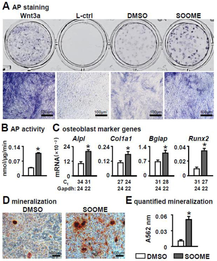
Figure 2.
Effects of Wnt signaling-activated osteocytes on osteogenic differentiation in stromal cells. MLO-Y4 was treated with 60 µm S24 for 24 h, then co-cultured with ST2 cells for 3 days, and osteogenic differentiation was evaluated by AP staining (A). Wnt3a conditioned medium and its L control medium were used as positive and negative controls, respectively. Lower images are enlargements of upper images; scale bar = 100 µm. (B) AP biochemical activity assay. (C) Expression of osteoblast marker genes as measured by qPCR. The * symbol indicates p < 0.05 by Student t-test; n = 3. (D,E) Mineralization assay of ST2 cells co-cultured with SOOME in growth medium for 3 days and in osteogenic medium for another 14 days. The osteogenic medium was changed every three days. The mineralized bone nodules were photographed after alizarin red S staining, followed by quantification assay. The * symbol indicates p < 0.05 versus DMSO group by t-test; n = 3. Scale bar = 100 µm. Ct, cycle number detected by qPCR machine for a relative amount of target genes.

