Figure 3.
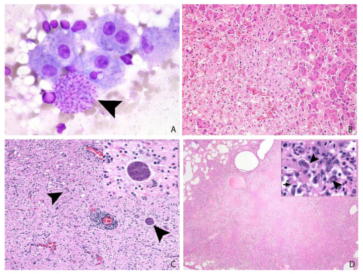
(A,B): Slender-tailed meerkat, 3 years, female, liver. A: Amongst hepatocytes, histiocytes and a background of erythrocytes there is a large cluster of intracytoplasmic Toxoplasma tachyzoites (arrow head); impression smear cytology, Leishman’s stain, ×100. (B): Large areas of coagulative necrosis efface hepatic parenchyma. Organisms are difficult to identify: H&E, ×20. (C): Pallas’ cat, 9 years, female, brain: Moderate to marked mixed mononuclear encephalitis with perivascular cuffing and intralesional protozoa. H&E, ×10, inset ×60. (D): Eurasian badger, adult, female, lung: Severe coalescent necrotising pneumonia. Intralesional intracellular tachyzoites (arrow heads) are present, and the cytoplasm of macrophages contain intralesional bacteria (arrow) (identified as mycobacteria on special stains). H&E, ×2, inset ×100.

