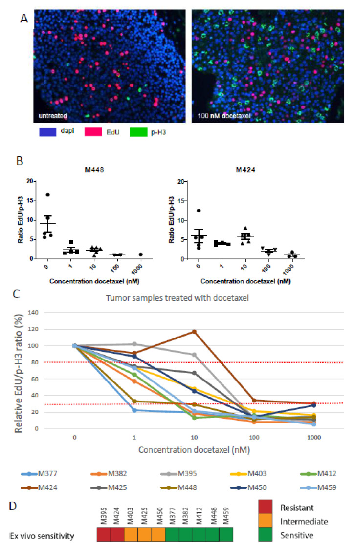
Figure 4.
Ex vivo docetaxel treatment of primary breast cancer. (A) Typical microscopic image of DAPI, EdU and p-H3 staining of primary BC slices without and with 3 days of 100 nM docetaxel treatment. (B) EdU/p-H3 ratios of two primary BC samples incubated for three days with various docetaxel concentrations. Each data point (circle, triangle or square) is the score for one microscopic field of view, with the mean and SEM indicated for each docetaxel concentration. (C) The EdU/p-H3 ratio in response to 3 days of incubation with the indicated docetaxel concentrations relative to the untreated control for 10 primary BC samples. Dotted red lines indicate the proposed thresholds for optimal discrimination between sensitive, intermediate and resistant tumors. (D) Overall results of ex vivo docetaxel sensitivity in the primary tumors using 30% and 80% of the relative EdU/p-H3 ratio at 10 nM docetaxel as thresholds for discriminating sensitive, intermediate and resistant tumors. M-numbers represent the individual primary mammary tumors.

