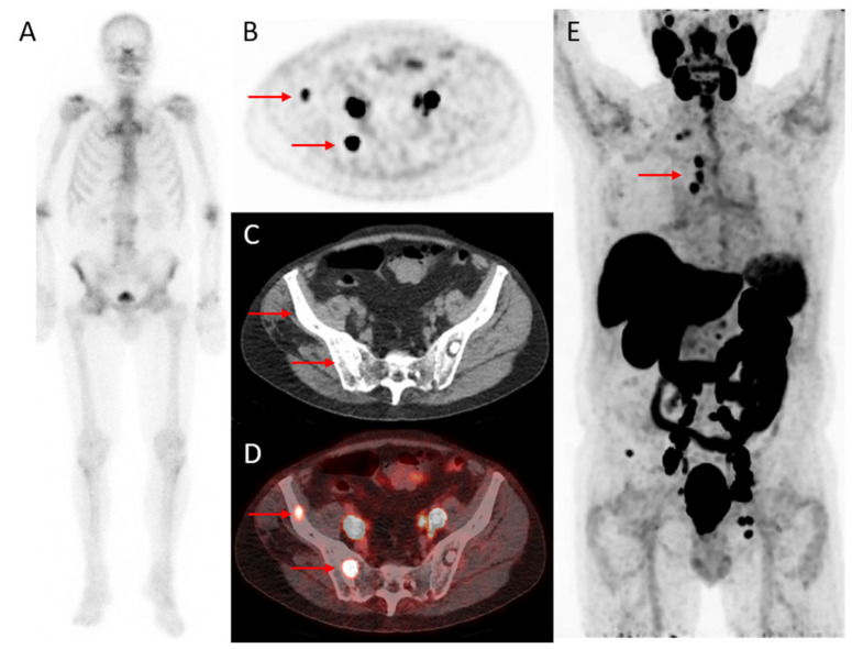
Figure 2.
A 79-year-old patient with CRPC after initial treatment with radiotherapy followed by hormonal therapy. Images illustrate improved detection of bone metastases using 18F-DCFPyL PET/CT compared to bone scintigraphy (4 weeks interval). The PSA level at PET was 23 ng/mL. On bone scintigraphy, faint uptake in the lumbar spine, the right acromioclavicular joint, the sternoclavicular, and hip joints were attributed to degenerative changes (A). Transversal 18F-DCFPyL PET (B) and fused PET/CT (D) revealed two foci (red arrows) with intense PSMA-expression in the right iliac bone (SUVmax: cranial lesion 6.2 and caudal lesion 17) and a sclerotic substrate on CT (C) and were classified as highly suspicious for bone metastases. Maximum intensity projection (E) demonstrated additional lymph node metastases above the diaphragm.

