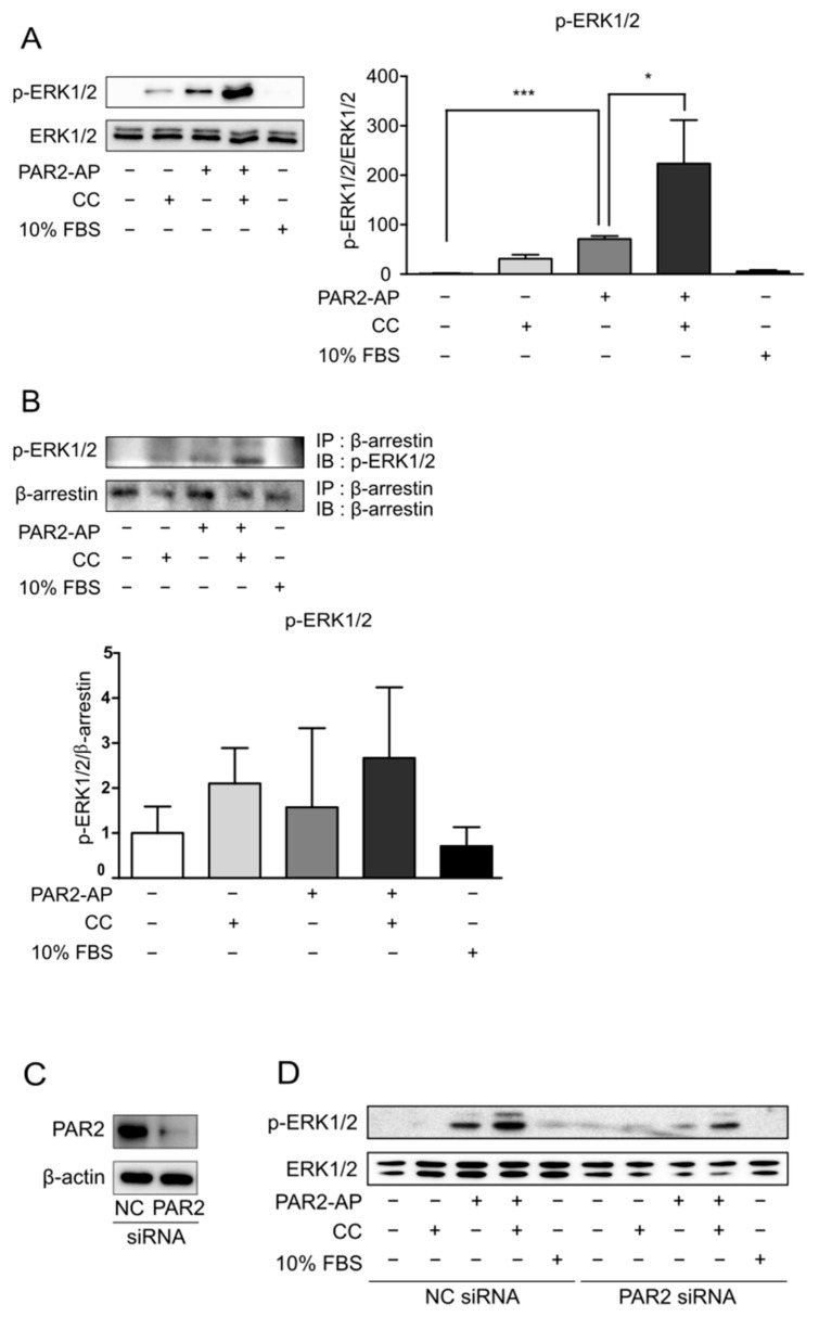
Figure 2.
PAR2 activation led to the recruitment of β-arrestin and ERK1/2. Cells were serum-starved for 24 h and treated with PAR2-AP (100 µM) or CC (IL-1β, 1 ng/mL; TNF-α, 20 ng/mL; and IFN-γ, 10 ng/mL) or 10% FBS for 6 h. (A) The protein level of p-ERK1/2 was measured using Western blotting. The bar graph indicates the ratio of p-ERK1/2 to ERK1/2. Data are shown as mean ± SD. * p < 0.05, *** p < 0.001 (one-way ANOVA). (B) Cell lysates were immunoprecipitated with β-arrestin antibody, followed by IB with p-ERK1/2 antibody. The bar graph indicates the ratio of p-ERK1/2 to β-arrestin. Data are shown as mean ± SD. Caco-2 cells were transfected with PAR2 siRNA for 48 h and then treated with PAR2-AP (100 µM) or CC (IL-1β, 1 ng/mL; TNF-α, 20 ng/mL; and IFN-γ, 10 ng/mL) or 10% FBS for 6 h. (C) Western blotting was used to detect a decrease in PAR2 protein expression in PAR2-KD cells. (D) p-ERK1/2 protein levels in PAR2-KD cells were measured using Western blotting.

