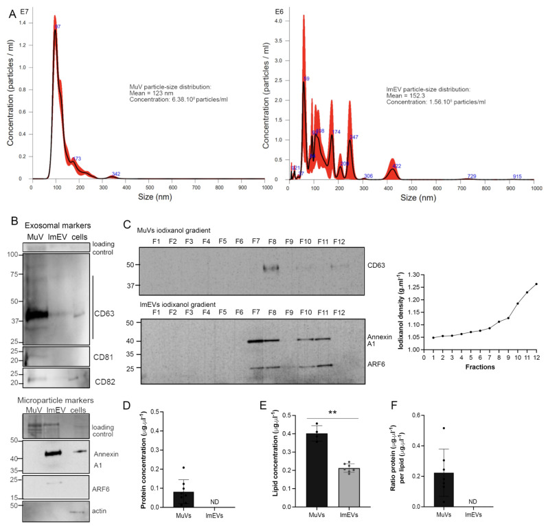
Figure 1.
MuVs and lmEVs present different markers and have different buoyant properties. (A) histograms showing the MuV and lmEV particle-size distributions (representative sample, from ALS EVs). (B) Representative Western blots showing the detection of CD63, CD81, CD82, AnnexinA1, ARF6, and actin, in MuVs (line1), lmEVs (line 2) and cells (line 3). Exosomal markers were enriched in MuVs and at relatively low or undetectable levels in lmEVs (EVs were extracted from the same cell culture medium for both exosomal and microparticle markers). Protein loaded on the gel is also shown, as loading control. Cellular contamination was not observed as neither of the vesicle fractions were positive for alpha-skeletal actin. (C) Vesicle extracts loaded on iodixanol gradients. MuVs presented classic exosomal buoyant properties while the buoyant range of lmEVs extended to a higher iodixanol density. Top panel: representative Western blot showing detection of CD63 for the MuVs at a density of 1.112 g·mL−1; bottom panel: representative Western blot (EVs extracted from the same cell culture medium as top panel) showing detection of Annexin A1 and ARF6 for the lmEVs at a density of 1.086–1.112 g·mL−1 and 1.185–1.230 g·mL−1. Right panel: iodixanol/sucrose density across the 12 fractions; gradient range from 1.048 to 1.263 g·mL−1. (D) Protein concentration in MuVs samples. Protein quantification was below detection sensitivity for lmEVs (ND: not detected). (E) Lipid quantification in MuVs and lmEVs. **, p < 0.01. (F) Ratio of protein per lipid in MuVs and lmEVs. The protein/lipid ratio could not be determined for lmEVs as the protein quantity was below detection sensitivity.

