Figure 2.
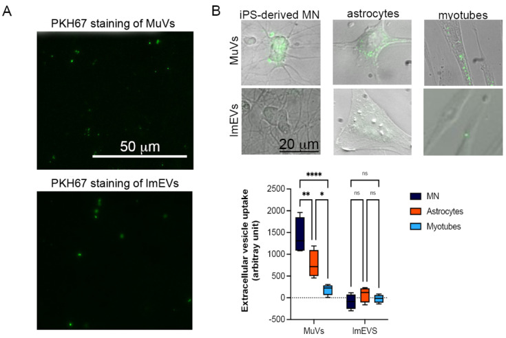
Uptake of MuVs or lmEVs by motor neurons, astrocytes, and myotubes. (A) MuVs and lmEVs labelled with PKH67 (droplet of vesicle preparations under fluorescence microscope), Bar = 50 μm. (B) A total of 85 ng of vesicular lipids were added to 10,000 cells. Top panel: representative images of MuV and lmEV uptake by hiPSC-derived motor neurons, astrocytes and myotubes. Vesicles were labeled with PKH67 (green). Bar = 25 μm. Bottom panel: the uptake of MuVs or lmEVs was assessed in healthy iPSC-derived motor neurons (MN), astrocytes, and myotubes (n = 4 per treatment, per cell line). Uptake of MuVs by MN was greater than by astrocytes or myotubes. Two-way ANOVA followed by Šídák’s multiple comparisons test. *, **, ****, p < 0.05, p < 0.01, p < 0.0001, respectively, ns: non-significant.

