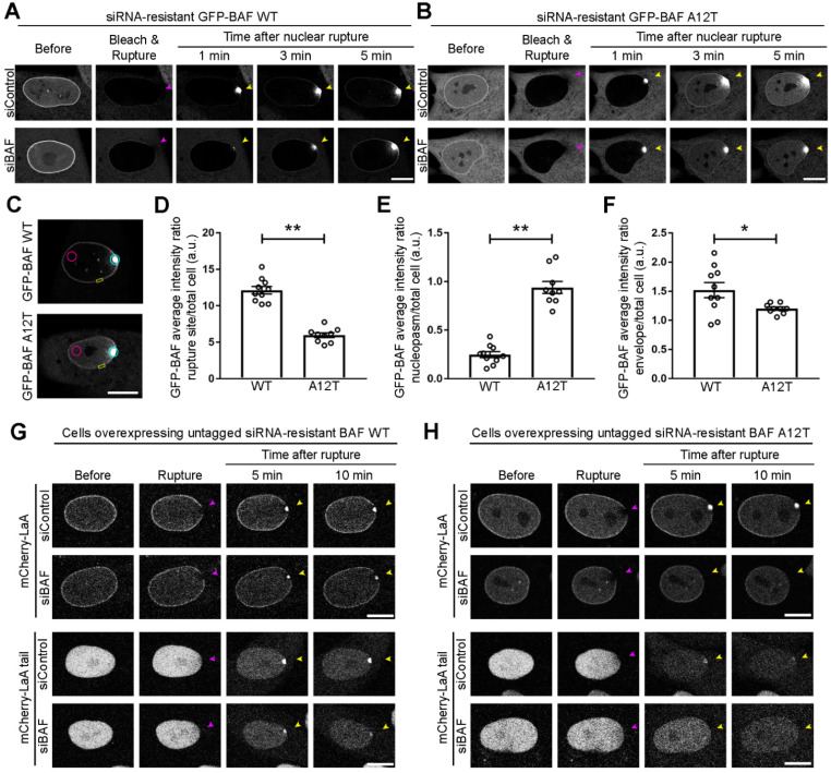
Figure 5.
Progeric BAF A12T exhibits reduced NE-association prior to and during rupture and is unable to recruit lamin A to nuclear ruptures. BJ-5ta cells expressing a GFP-tagged siRNA-resistant human BAF (A) WT or (B) A12T underwent endogenous BAF depletion via siRNA transfection prior to nuclear compartment photobleaching and laser-induced nuclear rupture. Migration of GFP-BAF into the nucleus was monitored for 5 min. Purple arrowheads indicate site of laser-induced rupture. Yellow arrowheads indicate location of expected protein accumulation. (C) Representative location of regions of interest (ROI) for quantification at 3 min post rupture of the average GFP-BAF intensity at the rupture site (blue ROI), nucleoplasm distal to rupture site (pink ROI), and nuclear envelope (NE) (yellow ROI). The average GFP-BAF intensity ratio of the (D) rupture site, (E) the nucleoplasm distal from the rupture site, or (F) a site on the nuclear envelope adjacent to the rupture was compared to average total cell intensity of GFP-BAF at 3 min post rupture. The graph represents mean values ± SEM and includes individual values (n = 10 cells for WT and 9 cells for A12T, ** p < 0.0001 and * p < 0.05 by an unpaired student’s t test). BJ-5ta cells stably co-expressing expressing GFP-NLS and untagged siRNA-resistant BAF (G) WT or (H) A12T via an internal ribosomal entry site (IRES) were co-expressed with either mCherry-LaA or mCherry-LaA tail and depleted of endogenous BAF via siRNA transfection prior to laser-induced nuclear rupture. Lamin accumulation at the rupture site was monitored over 10 min following rupture. Scale bars, 10 μm.

