Figure 2.
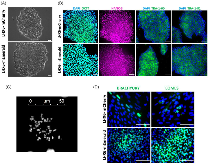
Reporter lines preserve pluripotent characteristics. (A) Phase contrast image of LHX6-mCherry and LHX6-mEmerald colonies showing characteristic hPSC morphology. (B) Double antibody staining for OCT4 and NANOG, and single staining for TRA 1–60 and TRA 1–81, respectively. (C) An example image of chromosome spread. (D) Immunostaining for Brachyury Y+ and EOMES in 15-day random differentiated LHE and LHM cultures. Scale bars in (A,C,D): 50 µ; (B): 100 µ.

