Figure 4.
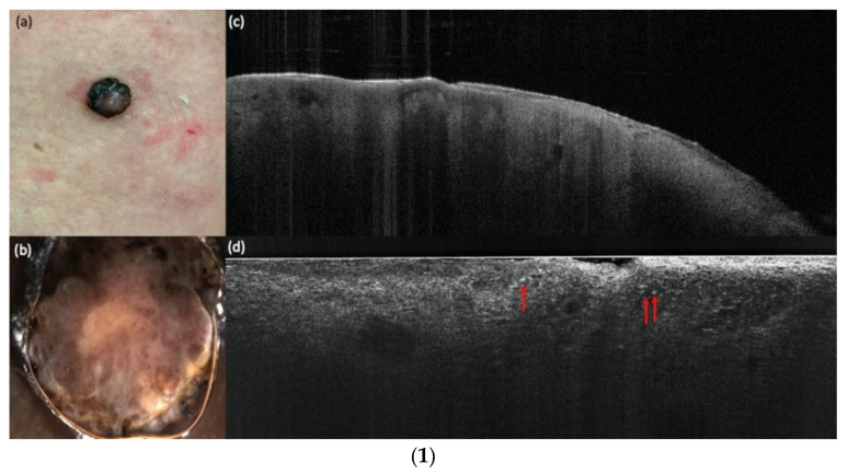
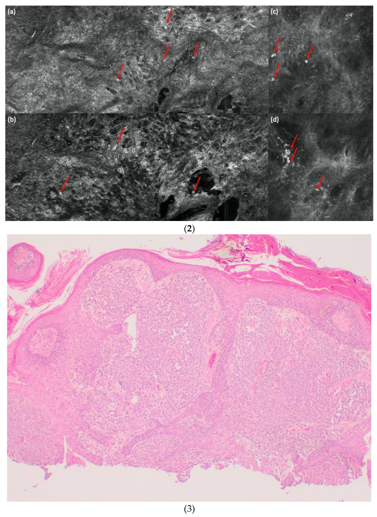
(1) A melanoma in comparison with vertical LC-OCT and OCT. An ulcerated nodular melanoma on the upper right leg. (a) Clinical, (b) dermoscopical, and (c) optical coherence tomography (OCT; 6 mm × 2 mm) images of a representative melanoma of the study. (d) In vertical line-field optical coherence tomography (LC-OCT; 1.2 mm × 0.5 mm) images, bright atypical melanocytic cells (red arrows) and a disturbed dermo-epidermal junction (DEJ) are visible. No dermal nests can be seen. (2) A melanoma in comparison with horizontal LC-OCT and RCM. The same melanoma is depicted in horizontal LC-OCT (1.2 mm × 0.5 mm) with an irregular honeycomb pattern with single atypical cells (red arrows) (a), and a pagetoid spread with atypical melanocytes (red arrows) (b). There were no dermal nests, a more chaotic structure, and no edged papillae visible. Reflectance confocal microscopy (RCM; 500 µm × 500 µm) shows the same features. An irregular honeycomb pattern with single atypical cells, just brighter (red arrows), (c) and a lighter pagetoid spread with atypical melanocytes (red arrows) (d). There were also no dermal nests, a more chaotic structure, and no edged papillae visible. (3) A melanoma in histology. The histology shows the same ulcerated nodular melanoma as in (1) and (2) with a tumor thickness of 4.8 mm and with 4× magnification.


