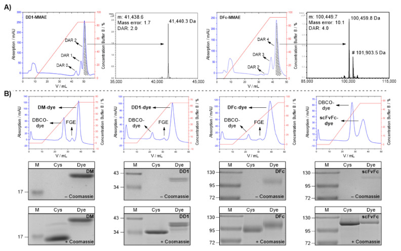
Figure 2.
Purification and analysis of protein-drug/dye conjugates. (A) Purification and analysis of DD1-MMAE (left) and DFc-MMAE (right) conjugates. HIC elution chromatogram from DD1 and DFc conjugated by tandem Knoevenagel-azide (7) and DBCO-PEG2-Lys(mPEG10)-βAla-Val-Cit-PAB-MMAE (6). Associated LC-ESI-ToF MS of the isolated MMAE conjugates (DD1-MMAE: 41,440.3 Da, DFc-MMAE: 100,459.8 Da). Residual glycosylated DFc-MMAE conjugate can be detected by signal shifted of 1443.7 Da (#). (B) Synthesis of Alexa Fluor 647 conjugates. Top: Hic elution chromatogram from the purification of the DM-, DD1-, DFc- and scFvFc-Alexa Fluor 647 conjugates. Middle: SDS PAGE analysis (with 5 mM DTT) of CTPSR-tagged proteins (Cys) and dye conjugates (Dye) before Coomassie staining. A pre-stained protein ladder (M) was used. The staining of the dye conjugate in the absence of Coomassie is caused by the color of the fluorophore. Bottom: SDS-PAGE analysis after Coomassie staining. Full SDS gels can be found in SI. (Figures S15–S18).

