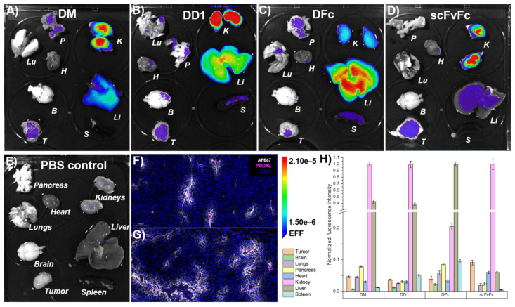
Figure 6.
Ex vivo imaging of anti-EGFR dye conjugates. A431-xenografted mice were intravenously injected with Alexa Fluor 647-labeled (A) DM, (B) DD1, (C) DFc, (D) scFvFc dye conjugates, or (E) PBS solution. Organs were collected following PFA-perfusion at 6 h post-injection. (F,G) Immunofluorescence staining showed Alexa Fluor 647 fluorescence (white) around tumor-associated podocalyxin (magenta) stained blood vessels of mice treated with DD1 (F) or scFvFc (G) -dye conjugates (n = 5 tumor sections, n = 1 tumor 6 h post-injection). Images represent a 20× magnification of a representative 10 µm tumor section taken by a slide scanner. (H) The mean signal intensity of Alexa Fluor 647 for each organ was quantified from ex vivo images using a 2 × 3 grid (n = 6) with Aura imaging software. Autofluorescence (from E) subtraction was separately done for each organ and resulting values were normalized to the highest fluorescence intensity among each set of organs (A–E). Error bars represent standard error of the mean. B: Brain, H: Heart, K: Kidneys, Li: Liver, Lu: Lungs, P: Pancreas, S: Spleen, T: Tumor.

